 Movie
Movie Controller


+ Open data

- Basic information
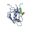
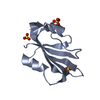
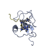
| Entry | Database: PDB / ID: 3ulj | ||||||
|---|---|---|---|---|---|---|---|
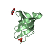
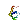
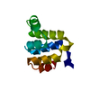
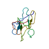
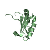
| Title | Crystal structure of apo Lin28B cold shock domain | ||||||
 Components Components | Lin28b, DNA-binding protein | ||||||
 Keywords Keywords | DNA BINDING PROTEIN / beta barrel / cold shock domain fold / nucleic acid binding | ||||||
| Function / homology |  Function and homology information Function and homology informationpre-miRNA processing / mRNA binding / nucleolus / zinc ion binding / nucleus / cytoplasm Similarity search - Function | ||||||
| Biological species | Xenopus tropicalis | ||||||
| Method |  X-RAY DIFFRACTION / SYNCHROTRON / MOLECULAR REPLACEMENT / Resolution: 1.06 Å X-RAY DIFFRACTION / SYNCHROTRON / MOLECULAR REPLACEMENT / Resolution: 1.06 Å | ||||||
 Authors Authors | Mayr, F. / Schuetz, A. / Doege, N. / Heinemann, U. | ||||||
 Citation Citation | Journal: Nucleic Acids Res. / Year: 2012 Title: The Lin28 cold-shock domain remodels pre-let-7 microRNA. Authors: Mayr, F. / Schutz, A. / Doge, N. / Heinemann, U. | ||||||
| History |
|
- Structure visualization
| Structure viewer | Molecule: MolmilJmol/JSmol |
|---|
- Downloads & links
-Download
| PDBx/mmCIF format | 3ulj.cif.gz | 100.3 KB | Display | PDBx/mmCIF format |
|---|---|---|---|---|
| PDB format | pdb3ulj.ent.gz | 78 KB | Display | PDB format |
| PDBx/mmJSON format | 3ulj.json.gz | Tree view | PDBx/mmJSON format | |
| Others |  Other downloads Other downloads |
-Validation report
| Arichive directory | https://data.pdbj.org/pub/pdb/validation_reports/ul/3uljftp://data.pdbj.org/pub/pdb/validation_reports/ul/3ulj | HTTPS FTP |
|---|
-Related structure data
| Related structure data |  4a4iC 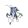 4a75C 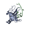 4a76C 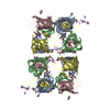 4alpC  312zS S: Starting model for refinement C: citing same article ( |
|---|---|
| Similar structure data |


-Links
 PDBj
PDBj- Assembly
| Deposited unit | 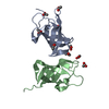
| ||||||||
|---|---|---|---|---|---|---|---|---|---|
| 1 | 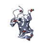
| ||||||||
| 2 | 
| ||||||||
| 3 | 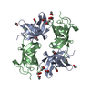
| ||||||||
| Unit cell |
|
-Components
| #1: Protein | Mass: 9942.231 Da / Num. of mol.: 2 / Fragment: unp residues 27-114 Source method: isolated from a genetically manipulated source Source: (gene. exp.) Gene: lin28b / Plasmid: pQLinkH / Production host:  #2: Chemical | ChemComp-ACT /   Mass: 59.044 Da / Num. of mol.: 8 / Source method: obtained synthetically / Formula: C2H3O2 Mass: 59.044 Da / Num. of mol.: 8 / Source method: obtained synthetically / Formula: C2H3O2#3: Chemical | ChemComp-GOL / |   Mass: 92.094 Da / Num. of mol.: 1 / Source method: obtained synthetically / Formula: C3H8O3 Mass: 92.094 Da / Num. of mol.: 1 / Source method: obtained synthetically / Formula: C3H8O3#4: Water | ChemComp-HOH / |  Mass: 18.015 Da / Num. of mol.: 272 / Source method: isolated from a natural source / Formula: H2O Mass: 18.015 Da / Num. of mol.: 272 / Source method: isolated from a natural source / Formula: H2O |
|---|
-Experimental details
-Experiment
| Experiment | Method: X-RAY DIFFRACTION / Number of used crystals: 1 |
|---|
- Sample preparation
| Crystal | Density Matthews: 2.76 Å3/Da / Density % sol: 55.29 % |
|---|---|
| Crystal grow | Method: evaporation / pH: 7 Details: 2.5 M sodium acetate, 0.1 M HEPES , pH 7.0, EVAPORATION |
-Data collection
| Diffraction | Mean temperature: 100 K |
|---|---|
| Diffraction source | Source: SYNCHROTRON / Site: BESSY  / Beamline: 14.1 / Wavelength: 0.98141 Å / Beamline: 14.1 / Wavelength: 0.98141 Å |
| Detector | Type: RAYONIX MX225HE / Detector: CCD / Date: Jul 29, 2010 |
| Radiation | Protocol: SINGLE WAVELENGTH / Monochromatic (M) / Laue (L): M / Scattering type: x-ray |
| Radiation wavelength | Wavelength: 0.98141 Å / Relative weight: 1 |
| Reflection | Resolution: 1.06→32.3 Å / Num. all: 100674 / Num. obs: 100170 / % possible obs: 99.5 % / Observed criterion σ(F): 2 / Observed criterion σ(I): 2 / Rmerge(I) obs: 0.056 / Rsym value: 0.056 / Net I/σ(I): 17.8 |
| Reflection shell | Resolution: 1.06→1.087 Å / Rmerge(I) obs: 0.61 / Mean I/σ(I) obs: 2.8 / Rsym value: 0.61 / % possible all: 98.3 |
- Processing
| Software |
| ||||||||||||||||||||||||||||||||||||||||||||||||||||||||||||||||||||||||||||||||||||||||||
|---|---|---|---|---|---|---|---|---|---|---|---|---|---|---|---|---|---|---|---|---|---|---|---|---|---|---|---|---|---|---|---|---|---|---|---|---|---|---|---|---|---|---|---|---|---|---|---|---|---|---|---|---|---|---|---|---|---|---|---|---|---|---|---|---|---|---|---|---|---|---|---|---|---|---|---|---|---|---|---|---|---|---|---|---|---|---|---|---|---|---|---|
| Refinement | Method to determine structure: MOLECULAR REPLACEMENT Starting model: pdb entry 312Z Resolution: 1.06→32.3 Å / Cor.coef. Fo:Fc: 0.983 / Cor.coef. Fo:Fc free: 0.977 / Occupancy max: 1 / Occupancy min: 0.12 / SU B: 0.56 / SU ML: 0.013 / Cross valid method: THROUGHOUT / σ(F): 0 / ESU R Free: 0.021 / Stereochemistry target values: MAXIMUM LIKELIHOOD Details: HYDROGENS HAVE BEEN ADDED IN THE RIDING POSITIONS U VALUES : REFINED INDIVIDUALLY
| ||||||||||||||||||||||||||||||||||||||||||||||||||||||||||||||||||||||||||||||||||||||||||
| Solvent computation | Ion probe radii: 0.8 Å / Shrinkage radii: 0.8 Å / VDW probe radii: 1.4 Å / Solvent model: BABINET MODEL WITH MASK | ||||||||||||||||||||||||||||||||||||||||||||||||||||||||||||||||||||||||||||||||||||||||||
| Displacement parameters | Biso max: 66.98 Å2 / Biso mean: 15.9469 Å2 / Biso min: 3.86 Å2
| ||||||||||||||||||||||||||||||||||||||||||||||||||||||||||||||||||||||||||||||||||||||||||
| Refinement step | Cycle: LAST / Resolution: 1.06→32.3 Å
| ||||||||||||||||||||||||||||||||||||||||||||||||||||||||||||||||||||||||||||||||||||||||||
| Refine LS restraints |
| ||||||||||||||||||||||||||||||||||||||||||||||||||||||||||||||||||||||||||||||||||||||||||
| LS refinement shell | Resolution: 1.06→1.087 Å / Total num. of bins used: 20
|
