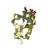
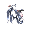
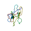
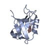
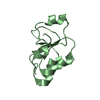
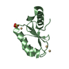
[English] 日本語
Yorodumi
- PDB-7d6f: The crystal structure of ARMS-PBM/MAGI2-PDZ4 -

+
Open data


ID or keywords:

Loading...

-
Basic information

Entry
Database: PDB / ID: 7d6f
TitleThe crystal structure of ARMS-PBM/MAGI2-PDZ4
Components
  • Kinase D-interacting substrate of 220 kDa
  • Membrane-associated guanylate kinase, WW and PDZ domain-containing protein 2
KeywordsSTRUCTURAL PROTEIN / PBM/PDZ interaction / scaffold protein / synaptic polaricity
Function / homology
Function and homology information


ARMS-mediated activation / structural constituent of postsynaptic specialization / extrinsic component of postsynaptic membrane / type II activin receptor binding / neuroligin clustering involved in postsynaptic membrane assembly / podocyte development / slit diaphragm / positive regulation of synaptic vesicle clustering / beta-1 adrenergic receptor binding / structural constituent of postsynaptic density ...ARMS-mediated activation / structural constituent of postsynaptic specialization / extrinsic component of postsynaptic membrane / type II activin receptor binding / neuroligin clustering involved in postsynaptic membrane assembly / podocyte development / slit diaphragm / positive regulation of synaptic vesicle clustering / beta-1 adrenergic receptor binding / structural constituent of postsynaptic density / nerve growth factor signaling pathway / activin receptor binding / protein kinase regulator activity / clathrin-dependent endocytosis / negative regulation of activin receptor signaling pathway / SMAD protein signal transduction / dendrite morphogenesis / regulation of neurotransmitter receptor localization to postsynaptic specialization membrane / regulation of synaptic membrane adhesion / ciliary base / receptor clustering / SMAD binding / positive regulation of phosphoprotein phosphatase activity / kinesin binding / positive regulation of receptor internalization / photoreceptor outer segment / negative regulation of phosphatidylinositol 3-kinase/protein kinase B signal transduction / GABA-ergic synapse / bicellular tight junction / phosphatase binding / photoreceptor inner segment / centriole / cellular response to nerve growth factor stimulus / negative regulation of cell migration / PDZ domain binding / positive regulation of neuron projection development / cell-cell junction / signaling receptor complex adaptor activity / late endosome / in utero embryonic development / postsynaptic density / membrane => GO:0016020 / negative regulation of cell population proliferation / centrosome / dendrite / glutamatergic synapse / synapse / protein-containing complex binding / protein kinase binding / perinuclear region of cytoplasm / signal transduction / protein-containing complex / nucleus / plasma membrane / cytosol / cytoplasm
Similarity search - Function
KAP family P-loop domain / KAP family P-loop domain / Unstructured region on MAGI / Domain of unknown function DUF3447 / Guanylate kinase, conserved site / Guanylate kinase-like signature. / Guanylate kinase-like domain profile. / Guanylate kinase-like domain / Guanylate kinase/L-type calcium channel beta subunit / Guanylate kinase ...KAP family P-loop domain / KAP family P-loop domain / Unstructured region on MAGI / Domain of unknown function DUF3447 / Guanylate kinase, conserved site / Guanylate kinase-like signature. / Guanylate kinase-like domain profile. / Guanylate kinase-like domain / Guanylate kinase/L-type calcium channel beta subunit / Guanylate kinase / Guanylate kinase homologues. / WW domain / WW/rsp5/WWP domain signature. / WW domain superfamily / WW/rsp5/WWP domain profile. / Domain with 2 conserved Trp (W) residues / WW domain / Sterile alpha motif/pointed domain superfamily / PDZ domain / PDZ domain profile. / Domain present in PSD-95, Dlg, and ZO-1/2. / PDZ domain / Ankyrin repeats (3 copies) / PDZ superfamily / Ankyrin repeat profile. / Ankyrin repeat region circular profile. / ankyrin repeats / Ankyrin repeat / Ankyrin repeat-containing domain superfamily / P-loop containing nucleoside triphosphate hydrolase
Similarity search - Domain/homology
Kinase D-interacting substrate of 220 kDa / Membrane-associated guanylate kinase, WW and PDZ domain-containing protein 2
Similarity search - Component
Biological speciesMus musculus (house mouse)
Rattus norvegicus (Norway rat)
MethodX-RAY DIFFRACTION / SYNCHROTRON / MOLECULAR REPLACEMENT / Resolution: 3.001 Å
AuthorsYe, J. / Zhang, Y. / Zhong, Z. / Wang, C.
CitationJournal: Neurochem.Int. / Year: 2021
Title: Crystal structure of the PDZ4 domain of MAGI2 in complex with PBM of ARMS reveals a canonical PDZ recognition mode.
Authors: Zhang, Y. / Zhong, Z. / Ye, J. / Wang, C.
History
DepositionSep 30, 2020Deposition site: PDBJ / Processing site: PDBJ
Revision 1.0Nov 4, 2020Provider: repository / Type: Initial release
Revision 2.0Sep 1, 2021Group: Advisory / Atomic model ...Advisory / Atomic model / Author supporting evidence / Data collection / Database references / Derived calculations / Non-polymer description / Refinement description / Structure summary
Category: atom_site / atom_site_anisotrop ...atom_site / atom_site_anisotrop / audit_author / chem_comp / citation_author / database_2 / entity / pdbx_entity_instance_feature / pdbx_entity_nonpoly / pdbx_entry_details / pdbx_nonpoly_scheme / pdbx_poly_seq_scheme / pdbx_refine_tls / pdbx_refine_tls_group / pdbx_struct_assembly_gen / pdbx_struct_assembly_prop / pdbx_struct_sheet_hbond / pdbx_validate_rmsd_angle / pdbx_validate_symm_contact / pdbx_validate_torsion / refine / refine_hist / refine_ls_shell / reflns / software / struct_asym / struct_conf / struct_sheet / struct_sheet_order / struct_sheet_range
Item: _audit_author.name / _chem_comp.formula ..._audit_author.name / _chem_comp.formula / _chem_comp.formula_weight / _chem_comp.id / _chem_comp.mon_nstd_flag / _chem_comp.name / _chem_comp.pdbx_synonyms / _chem_comp.type / _citation_author.name / _database_2.pdbx_DOI / _database_2.pdbx_database_accession / _pdbx_poly_seq_scheme.auth_mon_id / _pdbx_refine_tls.L[1][1] / _pdbx_refine_tls.L[1][2] / _pdbx_refine_tls.L[1][3] / _pdbx_refine_tls.L[2][2] / _pdbx_refine_tls.L[2][3] / _pdbx_refine_tls.L[3][3] / _pdbx_refine_tls.S[1][1] / _pdbx_refine_tls.S[1][2] / _pdbx_refine_tls.S[1][3] / _pdbx_refine_tls.S[2][1] / _pdbx_refine_tls.S[2][2] / _pdbx_refine_tls.S[2][3] / _pdbx_refine_tls.S[3][1] / _pdbx_refine_tls.S[3][2] / _pdbx_refine_tls.S[3][3] / _pdbx_refine_tls.T[1][1] / _pdbx_refine_tls.T[1][2] / _pdbx_refine_tls.T[1][3] / _pdbx_refine_tls.T[2][2] / _pdbx_refine_tls.T[2][3] / _pdbx_refine_tls.T[3][3] / _pdbx_refine_tls.origin_x / _pdbx_refine_tls.origin_y / _pdbx_refine_tls.origin_z / _pdbx_struct_assembly_gen.asym_id_list / _pdbx_struct_assembly_prop.value / _refine.B_iso_max / _refine.B_iso_mean / _refine.B_iso_min / _refine.ls_R_factor_R_free / _refine.ls_R_factor_R_work / _refine.ls_R_factor_obs / _refine.ls_d_res_high / _refine.ls_number_reflns_R_free / _refine.ls_number_reflns_R_work / _refine.ls_number_reflns_obs / _refine.ls_percent_reflns_R_free / _refine.ls_percent_reflns_obs / _refine.overall_SU_ML / _refine.pdbx_ls_sigma_F / _refine.pdbx_overall_phase_error / _refine.pdbx_starting_model / _refine_hist.d_res_high / _refine_hist.number_atoms_solvent / _refine_hist.number_atoms_total / _refine_hist.pdbx_B_iso_mean_ligand / _refine_hist.pdbx_B_iso_mean_solvent / _refine_hist.pdbx_number_atoms_ligand / _refine_ls_shell.R_factor_R_free / _refine_ls_shell.R_factor_R_work / _refine_ls_shell.d_res_high / _refine_ls_shell.d_res_low / _refine_ls_shell.number_reflns_R_free / _refine_ls_shell.number_reflns_R_work / _refine_ls_shell.percent_reflns_obs / _reflns.B_iso_Wilson_estimate / _reflns.d_resolution_high / _reflns.pdbx_number_measured_all / _software.version / _struct_conf.end_auth_comp_id / _struct_conf.end_auth_seq_id / _struct_conf.end_label_comp_id / _struct_conf.end_label_seq_id / _struct_conf.pdbx_PDB_helix_length / _struct_sheet.number_strands
Description: Real space R-factor
Details: To cut off the high resolution to 3.0 Angstrom when running the refinement program.
Provider: author / Type: Coordinate replacement
Revision 2.1Nov 17, 2021Group: Database references / Category: citation / citation_author
Item: _citation.country / _citation.journal_abbrev ..._citation.country / _citation.journal_abbrev / _citation.journal_id_CSD / _citation.journal_id_ISSN / _citation.journal_volume / _citation.page_first / _citation.page_last / _citation.pdbx_database_id_DOI / _citation.pdbx_database_id_PubMed / _citation.title / _citation.year / _citation_author.name
Revision 2.2Nov 29, 2023Group: Data collection / Refinement description
Category: chem_comp_atom / chem_comp_bond / pdbx_initial_refinement_model

-
Structure visualization

Structure viewerMolecule:
MolmilJmol/JSmol

Downloads & links

-
Assembly

Deposited unit
A: Membrane-associated guanylate kinase, WW and PDZ domain-containing protein 2
B: Kinase D-interacting substrate of 220 kDa
hetero molecules


Theoretical massNumber of molelcules
Total (without water)12,8123
Polymers12,7202
Non-polymers921
Water543
1


  • Idetical with deposited unit
  • defined by author&software
  • Evidence: light scattering
TypeNameSymmetry operationNumber
identity operation1_555x,y,z1
Buried area1000 Å2
ΔGint-7 kcal/mol
Surface area5240 Å2
MethodPISA
Unit cell
Length a, b, c (Å)74.045, 95.666, 35.167
Angle α, β, γ (deg.)90.000, 90.000, 90.000
Int Tables number21
Space group name H-MC222

-
Components

#1: Protein Membrane-associated guanylate kinase, WW and PDZ domain-containing protein 2 / / Activin receptor-interacting protein 1 / Acvrip1 / Atrophin-1-interacting protein 1 / AIP-1 / ...Activin receptor-interacting protein 1 / Acvrip1 / Atrophin-1-interacting protein 1 / AIP-1 / Membrane-associated guanylate kinase inverted 2 / MAGI-2


Mass: 10793.422 Da / Num. of mol.: 1
Source method: isolated from a genetically manipulated source
Source: (gene. exp.) Mus musculus (house mouse) / Gene: Magi2, Acvrinp1, Aip1, Arip1 / Production host: Escherichia coli (E. coli) / References: UniProt: Q9WVQ1
#2: Protein/peptide Kinase D-interacting substrate of 220 kDa / ARMS / Ankyrin repeat-rich membrane-spanning protein


Mass: 1926.969 Da / Num. of mol.: 1
Source method: isolated from a genetically manipulated source
Source: (gene. exp.) Rattus norvegicus (Norway rat) / Gene: Kidins220, Arms / Production host: Escherichia coli (E. coli) / References: UniProt: Q9EQG6
#3: Chemical ChemComp-GOL / GLYCEROL / GLYCERIN / PROPANE-1,2,3-TRIOL / Glycerol


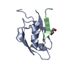
Mass: 92.094 Da / Num. of mol.: 1 / Source method: obtained synthetically / Formula: C3H8O3 / Feature type: SUBJECT OF INVESTIGATION
#4: Water ChemComp-HOH / water / Water


Mass: 18.015 Da / Num. of mol.: 3 / Source method: isolated from a natural source / Formula: H2O
Has ligand of interestY

-
Experimental details

-
Experiment

ExperimentMethod: X-RAY DIFFRACTION / Number of used crystals: 1

-
Sample preparation

CrystalDensity Matthews: 2.45 Å3/Da / Density % sol: 49.75 %
Crystal growTemperature: 277 K / Method: vapor diffusion, sitting drop
Details: 0.1 M HEPES pH 7.5, 10% w/v Polyethylene glycol 8000, 8% v/v Ethylene glycol

-
Data collection

DiffractionMean temperature: 100 K / Serial crystal experiment: N
Diffraction sourceSource: SYNCHROTRON / Site: SSRF / Beamline: BL19U1 / Wavelength: 0.97776 Å
DetectorType: DECTRIS EIGER X 16M / Detector: PIXEL / Date: May 8, 2017
RadiationProtocol: SINGLE WAVELENGTH / Monochromatic (M) / Laue (L): M / Scattering type: x-ray
Radiation wavelengthWavelength: 0.97776 Å / Relative weight: 1
ReflectionResolution: 2.7→50 Å / Num. obs: 3696 / % possible obs: 100 % / Redundancy: 12.5 % / Biso Wilson estimate: 83.67 Å2 / Rmerge(I) obs: 0.092 / Rpim(I) all: 0.027 / Rrim(I) all: 0.096 / Χ2: 0.684 / Net I/σ(I): 4.9 / Num. measured all: 46340
Reflection shell

Diffraction-ID: 1

Resolution (Å)Redundancy (%)Rmerge(I) obsNum. unique obsCC1/2Rpim(I) allRrim(I) allΧ2% possible all
2.7-2.7511.80.7241780.9670.2190.7570.485100
2.75-2.812.60.9741640.8680.2841.0150.491100
2.8-2.8512.20.6321820.9710.1850.6590.506100
2.85-2.9112.30.6941920.9520.2040.7240.51100
2.91-2.9713.30.6341700.9610.1790.660.484100
2.97-3.0413.40.5181880.9790.1440.5380.511100
3.04-3.1213.60.4251710.9730.1170.4410.53100
3.12-3.213.50.3132030.9930.0880.3250.528100
3.2-3.313.40.2511620.9930.070.2610.562100
3.3-3.412.90.1691860.9980.050.1770.614100
3.4-3.5213.40.1661850.9950.0470.1730.649100
3.52-3.6612.60.151770.9950.0440.1560.708100
3.66-3.8312.20.1091880.9980.0320.1140.765100
3.83-4.0311.30.1031850.9960.0310.1080.807100
4.03-4.2911.90.081800.9980.0240.0840.944100
4.29-4.6212.90.0731900.9980.0210.0761.095100
4.62-5.0812.90.0591970.9970.0170.0620.873100
5.08-5.8112.80.0611860.9980.0180.0640.797100
5.81-7.3211.50.0621940.9980.0190.0650.773100
7.32-5010.50.0472180.9990.0140.0491.05499.5

-
Processing

Software
NameVersionClassification
PHENIXv1.13refinement
HKL-2000data reduction
HKL-2000data scaling
PDB_EXTRACTdata extraction
MOLREP3.25phasing
RefinementMethod to determine structure: MOLECULAR REPLACEMENT
Starting model: 5ZYS
Resolution: 3.001→29.277 Å / SU ML: 0.11 / Cross valid method: THROUGHOUT / σ(F): 1.42 / Phase error: 20.16 / Stereochemistry target values: ML
RfactorNum. reflection% reflection
Rfree0.2814 137 5.09 %
Rwork0.2353 2552 -
obs0.2381 2689 99.63 %
Solvent computationShrinkage radii: 0.9 Å / VDW probe radii: 1.11 Å / Solvent model: FLAT BULK SOLVENT MODEL
Displacement parametersBiso max: 219.29 Å2 / Biso mean: 90.6272 Å2 / Biso min: 33.88 Å2
Refinement stepCycle: final / Resolution: 3.001→29.277 Å
ProteinNucleic acidLigandSolventTotal
Num. atoms657 0 6 3 666
Biso mean--134.18 63.16 -
Num. residues----96
LS refinement shellResolution: 3→3.61 Å / Rfactor Rfree error: 0
RfactorNum. reflection% reflection
Rfree0.2814 137 -
Rwork0.2353 2552 -
obs--100 %
Refinement TLS params.Method: refined / Origin x: -10.3215 Å / Origin y: -15.8526 Å / Origin z: 6.0889 Å
111213212223313233
T0.5429 Å20.0955 Å2-0.0938 Å2-0.5709 Å2-0.2705 Å2--0.6016 Å2
L13.9231 °211.3462 °2-3.7456 °2-6.8912 °2-4.0202 °2--1.1742 °2
S-0.6112 Å °-1.9994 Å °2.1955 Å °-0.0172 Å °0.1679 Å °0.8567 Å °0.1862 Å °0.7536 Å °0.0882 Å °
Refinement TLS group
IDRefine-IDRefine TLS-IDSelection detailsAuth asym-IDAuth seq-ID
1X-RAY DIFFRACTION1allA916 - 1008
2X-RAY DIFFRACTION1allB1754 - 1762
3X-RAY DIFFRACTION1allC1 - 3
4X-RAY DIFFRACTION1allD1

+
About Yorodumi

-
News

-
Feb 9, 2022. New format data for meta-information of EMDB entries

New format data for meta-information of EMDB entries

  • Version 3 of the EMDB header file is now the official format.
  • The previous official version 1.9 will be removed from the archive.

Related info.:EMDB header

External links:wwPDB to switch to version 3 of the EMDB data model

-
Aug 12, 2020. Covid-19 info

Covid-19 info

URL: https://pdbj.org/emnavi/covid19.php

New page: Covid-19 featured information page in EM Navigator.

Related info.:Covid-19 info / Mar 5, 2020. Novel coronavirus structure data

+
Mar 5, 2020. Novel coronavirus structure data

Novel coronavirus structure data

Related info.:Yorodumi Speices / Aug 12, 2020. Covid-19 info

External links:COVID-19 featured content - PDBj / Molecule of the Month (242):Coronavirus Proteases

+
Jan 31, 2019. EMDB accession codes are about to change! (news from PDBe EMDB page)

EMDB accession codes are about to change! (news from PDBe EMDB page)

  • The allocation of 4 digits for EMDB accession codes will soon come to an end. Whilst these codes will remain in use, new EMDB accession codes will include an additional digit and will expand incrementally as the available range of codes is exhausted. The current 4-digit format prefixed with “EMD-” (i.e. EMD-XXXX) will advance to a 5-digit format (i.e. EMD-XXXXX), and so on. It is currently estimated that the 4-digit codes will be depleted around Spring 2019, at which point the 5-digit format will come into force.
  • The EM Navigator/Yorodumi systems omit the EMD- prefix.

Related info.:Q: What is EMD? / ID/Accession-code notation in Yorodumi/EM Navigator

External links:EMDB Accession Codes are Changing Soon! / Contact to PDBj

+
Jul 12, 2017. Major update of PDB

Major update of PDB

  • wwPDB released updated PDB data conforming to the new PDBx/mmCIF dictionary.
  • This is a major update changing the version number from 4 to 5, and with Remediation, in which all the entries are updated.
  • In this update, many items about electron microscopy experimental information are reorganized (e.g. em_software).
  • Now, EM Navigator and Yorodumi are based on the updated data.

External links:wwPDB Remediation / Enriched Model Files Conforming to OneDep Data Standards Now Available in the PDB FTP Archive

-
Yorodumi

Thousand views of thousand structures

  • Yorodumi is a browser for structure data from EMDB, PDB, SASBDB, etc.
  • This page is also the successor to EM Navigator detail page, and also detail information page/front-end page for Omokage search.
  • The word "yorodu" (or yorozu) is an old Japanese word meaning "ten thousand". "mi" (miru) is to see.

Related info.:EMDB / PDB / SASBDB / Comparison of 3 databanks / Yorodumi Search / Aug 31, 2016. New EM Navigator & Yorodumi / Yorodumi Papers / Jmol/JSmol / Function and homology information / Changes in new EM Navigator and Yorodumi

Read more