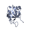
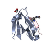
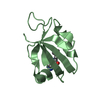
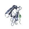
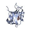
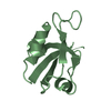
[English] 日本語
Yorodumi
- PDB-2aww: Synapse associated protein 97 PDZ2 domain variant C378G with C-te... -

+
Open data


ID or keywords:

Loading...

-
Basic information

Entry
Database: PDB / ID: 2aww
TitleSynapse associated protein 97 PDZ2 domain variant C378G with C-terminal GluR-A peptide
Components
  • 18-residue C-terminal peptide from glutamate receptor, ionotropic, AMPA1
  • Synapse associated protein 97
KeywordsMEMBRANE PROTEIN / synaptic signaling / trafficking protein
Function / homology
Function and homology information


tissue morphogenesis / L27 domain binding / regulation of protein localization to synapse / regulation of potassium ion import / MPP7-DLG1-LIN7 complex / regulation of potassium ion export across plasma membrane / membrane raft organization / Cargo concentration in the ER / axonal spine / positive regulation of locomotion involved in locomotory behavior ...tissue morphogenesis / L27 domain binding / regulation of protein localization to synapse / regulation of potassium ion import / MPP7-DLG1-LIN7 complex / regulation of potassium ion export across plasma membrane / membrane raft organization / Cargo concentration in the ER / axonal spine / positive regulation of locomotion involved in locomotory behavior / establishment of centrosome localization / myelin sheath abaxonal region / positive regulation of membrane potential / structural constituent of postsynaptic density / embryonic skeletal system morphogenesis / membrane repolarization during ventricular cardiac muscle cell action potential / COPII-mediated vesicle transport / epithelial structure maintenance / astral microtubule organization / reproductive structure development / immunological synapse formation / negative regulation of p38MAPK cascade / peristalsis / cellular response to ammonium ion / receptor localization to synapse / lateral loop / response to sucrose / smooth muscle tissue development / bicellular tight junction assembly / cell projection membrane / paranode region of axon / cortical microtubule organization / neuron spine / myosin V binding / protein localization to synapse / establishment or maintenance of epithelial cell apical/basal polarity / proximal dendrite / regulation of monoatomic ion transmembrane transport / regulation of ventricular cardiac muscle cell action potential / Trafficking of AMPA receptors / positive regulation of potassium ion transport / Activation of Ca-permeable Kainate Receptor / hard palate development / response to arsenic-containing substance / protein-containing complex localization / node of Ranvier / amyloid precursor protein metabolic process / cellular response to L-glutamate / cellular response to dsRNA / dendritic spine membrane / endothelial cell proliferation / long-term synaptic depression / lens development in camera-type eye / RAF/MAP kinase cascade / beta-2 adrenergic receptor binding / Synaptic adhesion-like molecules / cortical actin cytoskeleton organization / ureteric bud development / regulation of myelination / cellular response to peptide hormone stimulus / response to morphine / branching involved in ureteric bud morphogenesis / neuronal cell body membrane / protein kinase A binding / peptide hormone receptor binding / cellular response to amine stimulus / response to psychosocial stress / neurotransmitter receptor localization to postsynaptic specialization membrane / negative regulation of G1/S transition of mitotic cell cycle / spinal cord development / perisynaptic space / Activation of AMPA receptors / receptor clustering / positive regulation of actin filament polymerization / microvillus / AMPA glutamate receptor activity / response to lithium ion / Trafficking of GluR2-containing AMPA receptors / behavioral response to pain / AMPA glutamate receptor complex / basement membrane / adenylate cyclase binding / immunoglobulin binding / lateral plasma membrane / asymmetric synapse / immunological synapse / ionotropic glutamate receptor complex / excitatory synapse / conditioned place preference / response to electrical stimulus / regulation of receptor recycling / kinesin binding / G-protein alpha-subunit binding / glutamate receptor binding / bicellular tight junction / positive regulation of excitatory postsynaptic potential / Unblocking of NMDA receptors, glutamate binding and activation / long-term memory / positive regulation of synaptic transmission / potassium channel regulator activity
Similarity search - Function
L27-1 / L27_1 / domain in receptor targeting proteins Lin-2 and Lin-7 / L27 domain / L27 domain profile. / L27 domain superfamily / Polyubiquitination (PEST) N-terminal domain of MAGUK / Disks large homologue 1, N-terminal PEST domain / Polyubiquitination (PEST) N-terminal domain of MAGUK / PDZ-associated domain of NMDA receptors ...L27-1 / L27_1 / domain in receptor targeting proteins Lin-2 and Lin-7 / L27 domain / L27 domain profile. / L27 domain superfamily / Polyubiquitination (PEST) N-terminal domain of MAGUK / Disks large homologue 1, N-terminal PEST domain / Polyubiquitination (PEST) N-terminal domain of MAGUK / PDZ-associated domain of NMDA receptors / PDZ-associated domain of NMDA receptors / Disks large 1-like / : / Guanylate kinase, conserved site / Guanylate kinase-like signature. / Guanylate kinase-like domain profile. / Guanylate kinase-like domain / Guanylate kinase/L-type calcium channel beta subunit / Guanylate kinase / Guanylate kinase homologues. / PDZ domain / Pdz3 Domain / Ionotropic glutamate receptor, metazoa / Ligated ion channel L-glutamate- and glycine-binding site / Ionotropic glutamate receptor, L-glutamate and glycine-binding domain / Ligated ion channel L-glutamate- and glycine-binding site / Ligand-gated ion channel / : / Ionotropic glutamate receptor / Eukaryotic homologues of bacterial periplasmic substrate binding proteins. / PDZ domain / SH3 domain / Receptor, ligand binding region / Receptor family ligand binding region / PDZ domain profile. / Domain present in PSD-95, Dlg, and ZO-1/2. / PDZ domain / PDZ superfamily / Periplasmic binding protein-like I / Src homology 3 domains / SH3-like domain superfamily / Src homology 3 (SH3) domain profile. / SH3 domain / Roll / P-loop containing nucleoside triphosphate hydrolase / Mainly Beta
Similarity search - Domain/homology
: / Glutamate receptor 1 / Disks large homolog 1
Similarity search - Component
Biological speciesRattus norvegicus (Norway rat)
MethodX-RAY DIFFRACTION / SYNCHROTRON / MOLECULAR REPLACEMENT / Resolution: 2.21 Å
AuthorsVon Ossowski, I. / Oksanen, E. / Von Ossowski, L. / Cai, C. / Sundberg, M. / Goldman, A. / Keinanen, K.
CitationJournal: Febs J. / Year: 2006
Title: Crystal structure of the second PDZ domain of SAP97 in complex with a GluR-A C-terminal peptide
Authors: von Ossowski, I. / Oksanen, E. / von Ossowski, L. / Cai, C. / Sundberg, M. / Goldman, A. / Keinanen, K.
History
DepositionSep 2, 2005Deposition site: RCSB / Processing site: PDBJ
Revision 1.0Aug 29, 2006Provider: repository / Type: Initial release
Revision 1.1May 1, 2008Group: Version format compliance
Revision 1.2Jul 13, 2011Group: Version format compliance
Revision 1.3Oct 11, 2017Group: Refinement description / Category: software / Item: _software.classification / _software.name
Revision 1.4Nov 10, 2021Group: Database references / Category: database_2 / struct_ref_seq_dif
Item: _database_2.pdbx_DOI / _database_2.pdbx_database_accession / _struct_ref_seq_dif.details
Revision 1.5May 29, 2024Group: Data collection / Category: chem_comp_atom / chem_comp_bond

-
Structure visualization

Structure viewerMolecule:
MolmilJmol/JSmol

Downloads & links

-
Assembly

Deposited unit
A: Synapse associated protein 97
B: Synapse associated protein 97
C: 18-residue C-terminal peptide from glutamate receptor, ionotropic, AMPA1


Theoretical massNumber of molelcules
Total (without water)24,4853
Polymers24,4853
Non-polymers00
Water48627
1
A: Synapse associated protein 97
C: 18-residue C-terminal peptide from glutamate receptor, ionotropic, AMPA1


Theoretical massNumber of molelcules
Total (without water)13,1162
Polymers13,1162
Non-polymers00
Water181
TypeNameSymmetry operationNumber
identity operation1_555x,y,z1
Buried area610 Å2
ΔGint-2 kcal/mol
Surface area5150 Å2
MethodPISA
2
B: Synapse associated protein 97


Theoretical massNumber of molelcules
Total (without water)11,3691
Polymers11,3691
Non-polymers00
Water181
TypeNameSymmetry operationNumber
identity operation1_555x,y,z1
MethodPISA
Unit cell
Length a, b, c (Å)34.350, 54.310, 54.640
Angle α, β, γ (deg.)90.00, 84.03, 90.00
Int Tables number4
Space group name H-MP1211

-
Components

#1: Protein Synapse associated protein 97 / Presynaptic protein SAP97 / SAP-97


Mass: 11369.034 Da / Num. of mol.: 2 / Fragment: PDZ2 domain / Mutation: C378G
Source method: isolated from a genetically manipulated source
Source: (gene. exp.) Rattus norvegicus (Norway rat) / Gene: Dlg1 / Plasmid: PK0302-2 / Species (production host): Escherichia coli / Production host: Escherichia coli BL21(DE3) (bacteria) / Strain (production host): BL21 (DE3) / References: UniProt: Q62696
#2: Protein/peptide 18-residue C-terminal peptide from glutamate receptor, ionotropic, AMPA1 / 18-residue C-terminal peptide from GluR-A


Mass: 1747.048 Da / Num. of mol.: 1 / Source method: obtained synthetically / Details: Synthetic 18-residue peptide / References: GenBank: 29789269, UniProt: P19490*PLUS
#3: Water ChemComp-HOH / water


Mass: 18.015 Da / Num. of mol.: 27 / Source method: isolated from a natural source / Formula: H2O

-
Experimental details

-
Experiment

ExperimentMethod: X-RAY DIFFRACTION / Number of used crystals: 1

-
Sample preparation

CrystalDensity Matthews: 2.07 Å3/Da / Density % sol: 40.57 %
Crystal growTemperature: 293 K / Method: vapor diffusion, sitting drop / pH: 8.5
Details: 0.2M Sodium acetate, 0.1M Tris-HCl, 30% PEG4000, pH 8.5, VAPOR DIFFUSION, SITTING DROP, temperature 293K

-
Data collection

DiffractionMean temperature: 100 K
Diffraction sourceSource: SYNCHROTRON / Site: ESRF / Beamline: ID14-1 / Wavelength: 0.934 Å
DetectorType: ADSC QUANTUM 4 / Detector: CCD / Date: Nov 8, 2004
RadiationMonochromator: Diamond (111), germanium (220) / Protocol: SINGLE WAVELENGTH / Monochromatic (M) / Laue (L): M / Scattering type: x-ray
Radiation wavelengthWavelength: 0.934 Å / Relative weight: 1
ReflectionResolution: 2.2→50 Å / Num. all: 48906 / Num. obs: 48906 / % possible obs: 95.9 % / Observed criterion σ(F): 0 / Observed criterion σ(I): 0 / Rmerge(I) obs: 0.06 / Net I/σ(I): 17.3
Reflection shellResolution: 2.2→2.33 Å / % possible obs: 91.4 % / Rmerge(I) obs: 0.418 / Mean I/σ(I) obs: 3.78 / Num. measured obs: 7493 / Num. unique obs: 1485 / % possible all: 99.3

-
Phasing

Phasing MRRfactor: 0.488 / Cor.coef. Fo:Fc: 0.478
Highest resolutionLowest resolution
Rotation3 Å34.16 Å
Translation3 Å34.16 Å

-
Processing

Software
NameVersionClassificationNB
XSCALEdata scaling
MOLREPphasing
REFMAC5.2.0005refinement
PDB_EXTRACT1.7data extraction
ProDCdata collection
RefinementMethod to determine structure: MOLECULAR REPLACEMENT / Resolution: 2.21→34.1 Å / Cor.coef. Fo:Fc: 0.943 / Cor.coef. Fo:Fc free: 0.9 / SU B: 12.055 / SU ML: 0.293 / Cross valid method: THROUGHOUT / σ(F): 0 / ESU R: 0.305 / ESU R Free: 0.273 / Stereochemistry target values: MAXIMUM LIKELIHOOD / Details: HYDROGENS HAVE BEEN ADDED IN THE RIDING POSITIONS
RfactorNum. reflection% reflectionSelection details
Rfree0.326 509 5 %RANDOM
Rwork0.242 ---
all0.246 10167 --
obs0.246 10167 100 %-
Solvent computationIon probe radii: 0.8 Å / Shrinkage radii: 0.8 Å / VDW probe radii: 1.2 Å / Solvent model: MASK
Displacement parametersBiso mean: 48.035 Å2
Baniso -1Baniso -2Baniso -3
1-1.75 Å20 Å2-5.11 Å2
2---0.75 Å20 Å2
3---0.07 Å2
Refinement stepCycle: LAST / Resolution: 2.21→34.1 Å
ProteinNucleic acidLigandSolventTotal
Num. atoms1321 0 0 27 1348
Refine LS restraints
Refine-IDTypeDev idealDev ideal targetNumber
X-RAY DIFFRACTIONr_bond_refined_d0.0170.0221337
X-RAY DIFFRACTIONr_angle_refined_deg1.7521.981805
X-RAY DIFFRACTIONr_dihedral_angle_1_deg6.6765183
X-RAY DIFFRACTIONr_dihedral_angle_2_deg37.5127.04544
X-RAY DIFFRACTIONr_dihedral_angle_3_deg18.93815225
X-RAY DIFFRACTIONr_chiral_restr0.1050.2220
X-RAY DIFFRACTIONr_gen_planes_refined0.0060.02952
X-RAY DIFFRACTIONr_nbd_refined0.2370.2537
X-RAY DIFFRACTIONr_nbtor_refined0.3090.2887
X-RAY DIFFRACTIONr_xyhbond_nbd_refined0.2120.262
X-RAY DIFFRACTIONr_symmetry_vdw_refined0.2420.229
X-RAY DIFFRACTIONr_symmetry_hbond_refined0.0690.21
X-RAY DIFFRACTIONr_mcbond_it1.0171.5916
X-RAY DIFFRACTIONr_mcangle_it1.7621433
X-RAY DIFFRACTIONr_scbond_it2.2513449
X-RAY DIFFRACTIONr_scangle_it3.5344.5372
LS refinement shellResolution: 2.206→2.263 Å / Total num. of bins used: 20
RfactorNum. reflection% reflection
Rfree0.443 37 -
Rwork0.35 704 -
all-741 -
obs--100 %

+
About Yorodumi

-
News

-
Feb 9, 2022. New format data for meta-information of EMDB entries

New format data for meta-information of EMDB entries

  • Version 3 of the EMDB header file is now the official format.
  • The previous official version 1.9 will be removed from the archive.

Related info.:EMDB header

External links:wwPDB to switch to version 3 of the EMDB data model

-
Aug 12, 2020. Covid-19 info

Covid-19 info

URL: https://pdbj.org/emnavi/covid19.php

New page: Covid-19 featured information page in EM Navigator.

Related info.:Covid-19 info / Mar 5, 2020. Novel coronavirus structure data

+
Mar 5, 2020. Novel coronavirus structure data

Novel coronavirus structure data

Related info.:Yorodumi Speices / Aug 12, 2020. Covid-19 info

External links:COVID-19 featured content - PDBj / Molecule of the Month (242):Coronavirus Proteases

+
Jan 31, 2019. EMDB accession codes are about to change! (news from PDBe EMDB page)

EMDB accession codes are about to change! (news from PDBe EMDB page)

  • The allocation of 4 digits for EMDB accession codes will soon come to an end. Whilst these codes will remain in use, new EMDB accession codes will include an additional digit and will expand incrementally as the available range of codes is exhausted. The current 4-digit format prefixed with “EMD-” (i.e. EMD-XXXX) will advance to a 5-digit format (i.e. EMD-XXXXX), and so on. It is currently estimated that the 4-digit codes will be depleted around Spring 2019, at which point the 5-digit format will come into force.
  • The EM Navigator/Yorodumi systems omit the EMD- prefix.

Related info.:Q: What is EMD? / ID/Accession-code notation in Yorodumi/EM Navigator

External links:EMDB Accession Codes are Changing Soon! / Contact to PDBj

+
Jul 12, 2017. Major update of PDB

Major update of PDB

  • wwPDB released updated PDB data conforming to the new PDBx/mmCIF dictionary.
  • This is a major update changing the version number from 4 to 5, and with Remediation, in which all the entries are updated.
  • In this update, many items about electron microscopy experimental information are reorganized (e.g. em_software).
  • Now, EM Navigator and Yorodumi are based on the updated data.

External links:wwPDB Remediation / Enriched Model Files Conforming to OneDep Data Standards Now Available in the PDB FTP Archive

-
Yorodumi

Thousand views of thousand structures

  • Yorodumi is a browser for structure data from EMDB, PDB, SASBDB, etc.
  • This page is also the successor to EM Navigator detail page, and also detail information page/front-end page for Omokage search.
  • The word "yorodu" (or yorozu) is an old Japanese word meaning "ten thousand". "mi" (miru) is to see.

Related info.:EMDB / PDB / SASBDB / Comparison of 3 databanks / Yorodumi Search / Aug 31, 2016. New EM Navigator & Yorodumi / Yorodumi Papers / Jmol/JSmol / Function and homology information / Changes in new EM Navigator and Yorodumi

Read more