 Movie
Movie Controller

[English] 日本語
 Yorodumi
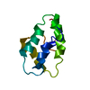
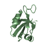
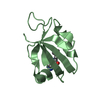
- PDB-2g2l: Crystal Structure of the Second PDZ Domain of SAP97 in Complex wi... -
+ Open data

- Basic information
| Entry | Database: PDB / ID: 2g2l | ||||||
|---|---|---|---|---|---|---|---|
| Title | Crystal Structure of the Second PDZ Domain of SAP97 in Complex with a GluR-A C-terminal Peptide | ||||||
 Components Components |
| ||||||
 Keywords Keywords | MEMBRANE PROTEIN / synaptic signaling / trafficking protein | ||||||
| Function / homology |  Function and homology information Function and homology informationregulation of signaling / tissue morphogenesis / L27 domain binding / regulation of protein localization to synapse / regulation of potassium ion import / MPP7-DLG1-LIN7 complex / regulation of potassium ion export across plasma membrane / membrane raft organization / establishment of centrosome localization / structural constituent of postsynaptic density ...regulation of signaling / tissue morphogenesis / L27 domain binding / regulation of protein localization to synapse / regulation of potassium ion import / MPP7-DLG1-LIN7 complex / regulation of potassium ion export across plasma membrane / membrane raft organization / establishment of centrosome localization / structural constituent of postsynaptic density / epithelial structure maintenance / reproductive structure development / membrane repolarization during ventricular cardiac muscle cell action potential / negative regulation of p38MAPK cascade / embryonic skeletal system morphogenesis / immunological synapse formation / receptor localization to synapse / myelin sheath abaxonal region / lateral loop / peristalsis / smooth muscle tissue development / cell projection membrane / paranode region of axon / cortical microtubule organization / bicellular tight junction assembly / astral microtubule organization / establishment or maintenance of epithelial cell apical/basal polarity / protein localization to synapse / positive regulation of potassium ion transport / Trafficking of AMPA receptors / hard palate development / Activation of Ca-permeable Kainate Receptor / regulation of ventricular cardiac muscle cell action potential / endothelial cell proliferation / amyloid precursor protein metabolic process / lens development in camera-type eye / ureteric bud development / node of Ranvier / cortical actin cytoskeleton organization / RAF/MAP kinase cascade / protein-containing complex localization / Synaptic adhesion-like molecules / regulation of myelination / branching involved in ureteric bud morphogenesis / negative regulation of G1/S transition of mitotic cell cycle / positive regulation of actin filament polymerization / neurotransmitter receptor localization to postsynaptic specialization membrane / receptor clustering / microvillus / AMPA glutamate receptor activity / basement membrane / lateral plasma membrane / AMPA glutamate receptor complex / immunological synapse / bicellular tight junction / potassium channel regulator activity / Unblocking of NMDA receptors, glutamate binding and activation / negative regulation of T cell proliferation / kinesin binding / regulation of postsynaptic membrane neurotransmitter receptor levels / phosphatase binding / postsynaptic density, intracellular component / response to fungicide / cellular response to brain-derived neurotrophic factor stimulus / presynaptic active zone membrane / T-tubule / ionotropic glutamate receptor binding / basal plasma membrane / negative regulation of phosphatidylinositol 3-kinase/protein kinase B signal transduction / protein localization to plasma membrane / actin filament organization / T cell activation / positive regulation of protein localization to plasma membrane / synaptic membrane / PDZ domain binding / neuromuscular junction / regulation of membrane potential / cell-cell adhesion / negative regulation of ERK1 and ERK2 cascade / cerebral cortex development / postsynaptic density membrane / negative regulation of epithelial cell proliferation / kinase binding / cytoplasmic side of plasma membrane / cell junction / cell-cell junction / intracellular protein localization / regulation of cell shape / nervous system development / regulation of protein localization / synaptic vesicle membrane / presynaptic membrane / chemical synaptic transmission / molecular adaptor activity / dendritic spine / basolateral plasma membrane / microtubule / transmembrane transporter binding / postsynaptic membrane / cell adhesion Similarity search - Function | ||||||
| Biological species |  | ||||||
| Method |  X-RAY DIFFRACTION / SYNCHROTRON / MOLECULAR REPLACEMENT / Resolution: 2.35 Å X-RAY DIFFRACTION / SYNCHROTRON / MOLECULAR REPLACEMENT / Resolution: 2.35 Å | ||||||
 Authors Authors | Von Ossowski, I. / Oksanen, E. / Von Ossowski, L. / Cai, C. / Sundberg, M. / Goldman, A. / Keinanen, K. | ||||||
 Citation Citation | Journal: Febs J. / Year: 2006 Title: Crystal structure of the second PDZ domain of SAP97 in complex with a GluR-A C-terminal peptide Authors: von Ossowski, I. / Oksanen, E. / von Ossowski, L. / Cai, C. / Sundberg, M. / Goldman, A. / Keinanen, K. | ||||||
| History |
|
- Structure visualization
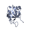
| Structure viewer | Molecule: MolmilJmol/JSmol |
|---|
- Downloads & links
-Download
| PDBx/mmCIF format | 2g2l.cif.gz | 47.9 KB | Display | PDBx/mmCIF format |
|---|---|---|---|---|
| PDB format | pdb2g2l.ent.gz | 33.4 KB | Display | PDB format |
| PDBx/mmJSON format | 2g2l.json.gz | Tree view | PDBx/mmJSON format | |
| Others |  Other downloads Other downloads |
-Validation report
| Arichive directory | https://data.pdbj.org/pub/pdb/validation_reports/g2/2g2lftp://data.pdbj.org/pub/pdb/validation_reports/g2/2g2l | HTTPS FTP |
|---|
-Related structure data
| Related structure data |  2awuC 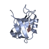 2awwC  2awxC  1pdrS C: citing same article ( S: Starting model for refinement |
|---|---|
| Similar structure data |


-Links
 PDBj
PDBj


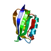
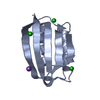
- Assembly
| Deposited unit | 
| ||||||||
|---|---|---|---|---|---|---|---|---|---|
| 1 | 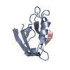
| ||||||||
| 2 | 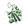
| ||||||||
| Unit cell |
|
-Components
| #1: Protein | Mass: 11471.255 Da / Num. of mol.: 2 / Fragment: PDZ2 domain Source method: isolated from a genetically manipulated source Source: (gene. exp.)  #2: Protein/peptide | Mass: 1747.048 Da / Num. of mol.: 2 / Source method: obtained synthetically / Details: synthetic 18-mer peptide / References: GenBank: 29789269, UniProt: M0R9A7*PLUS #3: Water | ChemComp-HOH / |  Mass: 18.015 Da / Num. of mol.: 44 / Source method: isolated from a natural source / Formula: H2O Mass: 18.015 Da / Num. of mol.: 44 / Source method: isolated from a natural source / Formula: H2O |
|---|
-Experimental details
-Experiment
| Experiment | Method: X-RAY DIFFRACTION / Number of used crystals: 1 |
|---|
- Sample preparation
| Crystal | Density Matthews: 1.97 Å3/Da / Density % sol: 37.5 % |
|---|---|
| Crystal grow | Temperature: 293 K / Method: vapor diffusion, sitting drop / pH: 8.5 Details: 0.2M SODIUM ACETATE, 0.1M TRIS-HCL, 30% PEG 4000 , pH 8.5, VAPOR DIFFUSION, SITTING DROP, temperature 293K |
-Data collection
| Diffraction | Mean temperature: 100 K |
|---|---|
| Diffraction source | Source: SYNCHROTRON / Site: ESRF  / Beamline: ID14-1 / Wavelength: 0.934 Å / Beamline: ID14-1 / Wavelength: 0.934 Å |
| Detector | Type: ADSC QUANTUM 4 / Detector: CCD / Date: Nov 8, 2004 |
| Radiation | Monochromator: DIAMOND (111) AND GE(220) / Protocol: SINGLE WAVELENGTH / Monochromatic (M) / Laue (L): M / Scattering type: x-ray |
| Radiation wavelength | Wavelength: 0.934 Å / Relative weight: 1 |
| Reflection | Resolution: 2.35→50 Å / Num. all: 8429 / Num. obs: 8429 / % possible obs: 97.5 % / Observed criterion σ(F): 0 / Observed criterion σ(I): 0 |
| Reflection shell | Resolution: 2.35→2.45 Å / % possible all: 96.6 |
- Processing
| Software |
| |||||||||||||||||||||||||||||||||||||||||||||||||||||||||||||||||||||||||||||||||||||
|---|---|---|---|---|---|---|---|---|---|---|---|---|---|---|---|---|---|---|---|---|---|---|---|---|---|---|---|---|---|---|---|---|---|---|---|---|---|---|---|---|---|---|---|---|---|---|---|---|---|---|---|---|---|---|---|---|---|---|---|---|---|---|---|---|---|---|---|---|---|---|---|---|---|---|---|---|---|---|---|---|---|---|---|---|---|---|
| Refinement | Method to determine structure: MOLECULAR REPLACEMENT Starting model: 1PDR Resolution: 2.35→38.63 Å / Cor.coef. Fo:Fc: 0.948 / Cor.coef. Fo:Fc free: 0.897 / SU B: 12.166 / SU ML: 0.276 / Cross valid method: THROUGHOUT / σ(F): 0 / ESU R: 0.383 / ESU R Free: 0.291 / Stereochemistry target values: MAXIMUM LIKELIHOOD / Details: HYDROGENS HAVE BEEN ADDED IN THE RIDING POSITIONS
| |||||||||||||||||||||||||||||||||||||||||||||||||||||||||||||||||||||||||||||||||||||
| Solvent computation | Ion probe radii: 0.8 Å / Shrinkage radii: 0.8 Å / VDW probe radii: 1.2 Å / Solvent model: MASK | |||||||||||||||||||||||||||||||||||||||||||||||||||||||||||||||||||||||||||||||||||||
| Displacement parameters | Biso mean: 45.075 Å2
| |||||||||||||||||||||||||||||||||||||||||||||||||||||||||||||||||||||||||||||||||||||
| Refinement step | Cycle: LAST / Resolution: 2.35→38.63 Å
| |||||||||||||||||||||||||||||||||||||||||||||||||||||||||||||||||||||||||||||||||||||
| Refine LS restraints |
| |||||||||||||||||||||||||||||||||||||||||||||||||||||||||||||||||||||||||||||||||||||
| LS refinement shell | Resolution: 2.353→2.414 Å / Total num. of bins used: 20
|
