 Movie
Movie Controller

[English] 日本語
 Yorodumi
- PDB-1pdr: CRYSTAL STRUCTURE OF THE THIRD PDZ DOMAIN FROM THE HUMAN HOMOLOG ... -
+ Open data

- Basic information
| Entry | Database: PDB / ID: 1pdr | ||||||
|---|---|---|---|---|---|---|---|
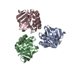
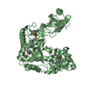
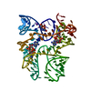
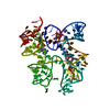
| Title | CRYSTAL STRUCTURE OF THE THIRD PDZ DOMAIN FROM THE HUMAN HOMOLOG OF DISCS LARGE PROTEIN | ||||||
 Components Components | HUMAN DISCS LARGE PROTEIN | ||||||
 Keywords Keywords | SIGNAL TRANSDUCTION / SH3 DOMAIN | ||||||
| Function / homology |  Function and homology information Function and homology informationL27 domain binding / regulation of protein localization to synapse / regulation of potassium ion import / MPP7-DLG1-LIN7 complex / regulation of potassium ion export across plasma membrane / membrane raft organization / establishment of centrosome localization / NrCAM interactions / GMP kinase activity / structural constituent of postsynaptic density ...L27 domain binding / regulation of protein localization to synapse / regulation of potassium ion import / MPP7-DLG1-LIN7 complex / regulation of potassium ion export across plasma membrane / membrane raft organization / establishment of centrosome localization / NrCAM interactions / GMP kinase activity / structural constituent of postsynaptic density / reproductive structure development / membrane repolarization during ventricular cardiac muscle cell action potential / negative regulation of p38MAPK cascade / embryonic skeletal system morphogenesis / immunological synapse formation / receptor localization to synapse / myelin sheath abaxonal region / lateral loop / peristalsis / smooth muscle tissue development / cell projection membrane / cortical microtubule organization / bicellular tight junction assembly / astral microtubule organization / establishment or maintenance of epithelial cell apical/basal polarity / regulation of sodium ion transmembrane transport / Synaptic adhesion-like molecules / protein localization to synapse / Trafficking of AMPA receptors / positive regulation of potassium ion transport / hard palate development / regulation of ventricular cardiac muscle cell action potential / endothelial cell proliferation / amyloid precursor protein metabolic process / lens development in camera-type eye / node of Ranvier / cortical actin cytoskeleton organization / Assembly and cell surface presentation of NMDA receptors / T cell proliferation / protein-containing complex localization / regulation of myelination / branching involved in ureteric bud morphogenesis / Activation of Ca-permeable Kainate Receptor / negative regulation of G1/S transition of mitotic cell cycle / positive regulation of actin filament polymerization / neurotransmitter receptor localization to postsynaptic specialization membrane / receptor clustering / establishment or maintenance of cell polarity / Negative regulation of NMDA receptor-mediated neuronal transmission / Unblocking of NMDA receptors, glutamate binding and activation / basement membrane / lateral plasma membrane / intercalated disc / immunological synapse / Long-term potentiation / bicellular tight junction / potassium channel regulator activity / negative regulation of T cell proliferation / regulation of postsynaptic membrane neurotransmitter receptor levels / phosphatase binding / cytoskeletal protein binding / phosphoprotein phosphatase activity / actin filament polymerization / ionotropic glutamate receptor binding / Ras activation upon Ca2+ influx through NMDA receptor / negative regulation of phosphatidylinositol 3-kinase/protein kinase B signal transduction / protein localization to plasma membrane / actin filament organization / positive regulation of protein localization to plasma membrane / synaptic membrane / adherens junction / phosphatidylinositol 3-kinase/protein kinase B signal transduction / neuromuscular junction / regulation of membrane potential / cell-cell adhesion / negative regulation of ERK1 and ERK2 cascade / sarcolemma / postsynaptic density membrane / negative regulation of epithelial cell proliferation / kinase binding / cytoplasmic side of plasma membrane / cell junction / cell-cell junction / regulation of cell shape / nervous system development / RAF/MAP kinase cascade / chemical synaptic transmission / molecular adaptor activity / basolateral plasma membrane / microtubule / transmembrane transporter binding / apical plasma membrane / neuron projection / cadherin binding / membrane raft / positive regulation of cell population proliferation / protein kinase binding / endoplasmic reticulum membrane / perinuclear region of cytoplasm / glutamatergic synapse Similarity search - Function | ||||||
| Biological species |  Homo sapiens (human) Homo sapiens (human) | ||||||
| Method |  X-RAY DIFFRACTION / Resolution: 2.8 Å X-RAY DIFFRACTION / Resolution: 2.8 Å | ||||||
 Authors Authors | Cabral, J.M. / Liddington, R. | ||||||
 Citation Citation | Journal: Nature / Year: 1996 Title: Crystal structure of a PDZ domain. Authors: Morais Cabral, J.H. / Petosa, C. / Sutcliffe, M.J. / Raza, S. / Byron, O. / Poy, F. / Marfatia, S.M. / Chishti, A.H. / Liddington, R.C. #1: Journal: Cell(Cambridge,Mass.) / Year: 1996Title: Clustering Membrane Proteins: It'S All Coming Together with the Psd-95/Sap90 Protein Family Authors: Gomperts, S.N. #2: Journal: Trends Biochem.Sci. / Year: 1995Title: Dhr Domains in Syntrophins, Neuronal No Synthases and Other Intracellular Proteins Authors: Ponting, C.P. / Phillips, C. #3: Journal: Nature / Year: 1995Title: Clustering of Shaker-Type K+ Channels by Interaction with a Family of Membrane-Associated Guanylate Kinases Authors: Kim, E. / Niethammer, M. / Rothschild, A. / Jan, Y.N. / Sheng, M. | ||||||
| History |
|
- Structure visualization
| Structure viewer | Molecule: MolmilJmol/JSmol |
|---|
- Downloads & links
-Download
| PDBx/mmCIF format | 1pdr.cif.gz | 32.6 KB | Display | PDBx/mmCIF format |
|---|---|---|---|---|
| PDB format | pdb1pdr.ent.gz | 22.2 KB | Display | PDB format |
| PDBx/mmJSON format | 1pdr.json.gz | Tree view | PDBx/mmJSON format | |
| Others |  Other downloads Other downloads |
-Validation report
| Arichive directory | https://data.pdbj.org/pub/pdb/validation_reports/pd/1pdrftp://data.pdbj.org/pub/pdb/validation_reports/pd/1pdr | HTTPS FTP |
|---|
-Related structure data
| Similar structure data |
|---|


-Links
 PDBj
PDBj


- Assembly
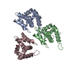
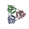
| Deposited unit | 
| ||||||||
|---|---|---|---|---|---|---|---|---|---|
| 1 | x 6
| ||||||||
| Unit cell |
|
-Components
| #1: Protein | Mass: 10556.708 Da / Num. of mol.: 1 / Fragment: PDZ3 DOMAIN, DHR3 DOMAIN Source method: isolated from a genetically manipulated source Details: THE THIRD PDZ DOMAIN OF THE HUMAN HOMOLOGUE OF DISCS LARGE PROTEIN Source: (gene. exp.) Homo sapiens (human) / Plasmid: PGEX-2T / Production host:  |
|---|---|
| #2: Water | ChemComp-HOH /  Mass: 18.015 Da / Num. of mol.: 8 / Source method: isolated from a natural source / Formula: H2O Mass: 18.015 Da / Num. of mol.: 8 / Source method: isolated from a natural source / Formula: H2O |
-Experimental details
-Experiment
| Experiment | Method: X-RAY DIFFRACTION |
|---|
- Sample preparation
| Crystal | Density Matthews: 5.26 Å3/Da / Density % sol: 76.61 % | |||||||||||||||
|---|---|---|---|---|---|---|---|---|---|---|---|---|---|---|---|---|
| Crystal grow | *PLUS Temperature: 4 ℃ / Method: vapor diffusion, hanging drop / PH range low: 7.5 / PH range high: 6.5 | |||||||||||||||
| Components of the solutions | *PLUS
|
-Data collection
| Diffraction source | Wavelength: 1.5418 |
|---|---|
| Detector | Type: RIGAKU / Detector: IMAGE PLATE / Date: Apr 12, 1996 |
| Radiation | Monochromatic (M) / Laue (L): M / Scattering type: x-ray |
| Radiation wavelength | Wavelength: 1.5418 Å / Relative weight: 1 |
| Reflection | Num. obs: 5911 / % possible obs: 100 % / Observed criterion σ(I): 0 / Redundancy: 13 % / Rmerge(I) obs: 0.108 |
| Reflection | *PLUS Highest resolution: 2.8 Å |
| Reflection shell | *PLUS Rmerge(I) obs: 0.301 |
- Processing
| Software |
| |||||||||||||||
|---|---|---|---|---|---|---|---|---|---|---|---|---|---|---|---|---|
| Refinement | Resolution: 2.8→10 Å / σ(F): 2 Details: SER 517 IS POSITIONED IN A VERY TIGHT LOOP AND THERE IS CLEAR DENSITY FOR ITS MAIN CHAIN ATOMS. THE PHI AND PSI ANGLES FOR THIS RESIDUE ARE OUTSIDE THE EXPECTED RANGE. THE DEPOSITORS ...Details: SER 517 IS POSITIONED IN A VERY TIGHT LOOP AND THERE IS CLEAR DENSITY FOR ITS MAIN CHAIN ATOMS. THE PHI AND PSI ANGLES FOR THIS RESIDUE ARE OUTSIDE THE EXPECTED RANGE. THE DEPOSITORS ATTEMPTED REPEATEDLY TO REGULARIZE THEM BUT THEY ALWAYS REFINED TO THE SAME VALUES.
| |||||||||||||||
| Displacement parameters | Biso mean: 16.7 Å2 | |||||||||||||||
| Refinement step | Cycle: LAST / Resolution: 2.8→10 Å
| |||||||||||||||
| Software | *PLUS Name: X-PLOR / Classification: refinement | |||||||||||||||
| Refinement | *PLUS | |||||||||||||||
| Solvent computation | *PLUS | |||||||||||||||
| Displacement parameters | *PLUS | |||||||||||||||
| Refine LS restraints | *PLUS
|
