[English] 日本語
Yorodumi
- PDB-4b8n: Cytochrome b5 of Ostreococcus tauri virus 2 -

+
Open data


ID or keywords:

Loading...

-
Basic information

Entry
Database: PDB / ID: 4b8n
TitleCytochrome b5 of Ostreococcus tauri virus 2
ComponentsCYTOCHROME B5-HOST ORIGIN
KeywordsELECTRON TRANSPORT / VIRAL CYTOCHROME B5
Function / homology
Function and homology information


heme binding / metal ion binding
Similarity search - Function
Flavocytochrome B2; Chain A, domain 1 / Cytochrome b5-like heme/steroid binding domain / Cytochrome b5, heme-binding site / Cytochrome b5 family, heme-binding domain signature. / Cytochrome b5 family, heme-binding domain profile. / Cytochrome b5-like heme/steroid binding domain / Cytochrome b5-like heme/steroid binding domain superfamily / Cytochrome b5-like Heme/Steroid binding domain / Cytochrome b5-like Heme/Steroid binding domain / Roll / Alpha Beta
Similarity search - Domain/homology
PROTOPORPHYRIN IX CONTAINING FE / Cytochrome b5-host origin
Similarity search - Component
Biological speciesOSTREOCOCCUS TAURI VIRUS 2
MethodX-RAY DIFFRACTION / SYNCHROTRON / MOLECULAR REPLACEMENT / Resolution: 1.95 Å
AuthorsIsupov, M. / Reid, E.L. / Weynberg, K.D. / Love, J. / Wilson, W.H. / Kelly, S.L. / Lamb, D.C. / Allen, M.J. / Littlechild, J.A.
CitationJournal: FEBS Lett. / Year: 2013
Title: Functional and Structural Characterisation of a Viral Cytochrome B5.
Authors: Reid, E.L. / Weynberg, K.D. / Love, J. / Isupov, M.N. / Littlechild, J.A. / Wilson, W.H. / Kelly, S.L. / Lamb, D.C. / Allen, M.J.
History
DepositionAug 28, 2012Deposition site: PDBE / Processing site: PDBE
Revision 1.0Sep 4, 2013Provider: repository / Type: Initial release
Revision 1.1Apr 23, 2014Group: Database references
Revision 1.2Dec 20, 2023Group: Data collection / Database references ...Data collection / Database references / Derived calculations / Other / Refinement description
Category: chem_comp_atom / chem_comp_bond ...chem_comp_atom / chem_comp_bond / database_2 / pdbx_database_status / pdbx_initial_refinement_model / pdbx_struct_conn_angle / struct_conn / struct_site
Item: _database_2.pdbx_DOI / _database_2.pdbx_database_accession ..._database_2.pdbx_DOI / _database_2.pdbx_database_accession / _pdbx_database_status.status_code_sf / _pdbx_struct_conn_angle.ptnr1_auth_seq_id / _pdbx_struct_conn_angle.ptnr1_label_seq_id / _pdbx_struct_conn_angle.ptnr3_auth_seq_id / _pdbx_struct_conn_angle.ptnr3_label_seq_id / _pdbx_struct_conn_angle.value / _struct_conn.pdbx_dist_value / _struct_conn.ptnr1_auth_comp_id / _struct_conn.ptnr1_auth_seq_id / _struct_conn.ptnr1_label_asym_id / _struct_conn.ptnr1_label_atom_id / _struct_conn.ptnr1_label_comp_id / _struct_conn.ptnr1_label_seq_id / _struct_conn.ptnr2_auth_comp_id / _struct_conn.ptnr2_auth_seq_id / _struct_conn.ptnr2_label_asym_id / _struct_conn.ptnr2_label_atom_id / _struct_conn.ptnr2_label_comp_id / _struct_conn.ptnr2_label_seq_id / _struct_site.pdbx_auth_asym_id / _struct_site.pdbx_auth_comp_id / _struct_site.pdbx_auth_seq_id

-
Structure visualization

Structure viewerMolecule:
MolmilJmol/JSmol

Downloads & links

-
Assembly

Deposited unit
A: CYTOCHROME B5-HOST ORIGIN
B: CYTOCHROME B5-HOST ORIGIN
C: CYTOCHROME B5-HOST ORIGIN
D: CYTOCHROME B5-HOST ORIGIN
hetero molecules


Theoretical massNumber of molelcules
Total (without water)44,48712
Polymers41,6374
Non-polymers2,8508
Water3,585199
1
A: CYTOCHROME B5-HOST ORIGIN
hetero molecules


Theoretical massNumber of molelcules
Total (without water)11,1223
Polymers10,4091
Non-polymers7132
Water181
TypeNameSymmetry operationNumber
identity operation1_555x,y,z1
MethodPISA
2
B: CYTOCHROME B5-HOST ORIGIN
hetero molecules


Theoretical massNumber of molelcules
Total (without water)11,1223
Polymers10,4091
Non-polymers7132
Water181
TypeNameSymmetry operationNumber
identity operation1_555x,y,z1
MethodPISA
3
C: CYTOCHROME B5-HOST ORIGIN
hetero molecules


Theoretical massNumber of molelcules
Total (without water)11,1223
Polymers10,4091
Non-polymers7132
Water181
TypeNameSymmetry operationNumber
identity operation1_555x,y,z1
MethodPISA
4
D: CYTOCHROME B5-HOST ORIGIN
hetero molecules


Theoretical massNumber of molelcules
Total (without water)11,1223
Polymers10,4091
Non-polymers7132
Water181
TypeNameSymmetry operationNumber
identity operation1_555x,y,z1
MethodPISA
Unit cell
Length a, b, c (Å)45.690, 58.500, 127.070
Angle α, β, γ (deg.)90.00, 90.00, 90.00
Int Tables number19
Space group name H-MP212121
Noncrystallographic symmetry (NCS)NCS oper:
IDCodeMatrixVector
1given(-0.417, -0.884, 0.211), (-0.908, 0.393, -0.145), (0.045, -0.252, -0.967)10.30088, 11.43419, -14.80462
2given(-0.27576, 0.96077, 0.02972), (0.96109, 0.27507, 0.02537), (0.0162, 0.03556, -0.99924)6.33218, 42.04393, -31.92822
3given(0.78907, -0.55908, 0.25455), (0.60425, 0.78104, -0.15766), (-0.11067, 0.27821, 0.95412)8.14446, 3.20634, 45.81707

-
Components

#1: Protein
CYTOCHROME B5-HOST ORIGIN


Mass: 10409.128 Da / Num. of mol.: 4
Source method: isolated from a genetically manipulated source
Source: (gene. exp.) OSTREOCOCCUS TAURI VIRUS 2 / Production host: ESCHERICHIA COLI (E. coli) / Strain (production host): BL21 / References: UniProt: E4WM77
#2: Chemical
ChemComp-HEM / PROTOPORPHYRIN IX CONTAINING FE / HEME


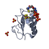
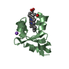
Mass: 616.487 Da / Num. of mol.: 4 / Source method: obtained synthetically / Formula: C34H32FeN4O4
#3: Chemical
ChemComp-SO4 / SULFATE ION


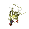
Mass: 96.063 Da / Num. of mol.: 4 / Source method: obtained synthetically / Formula: SO4
#4: Water ChemComp-HOH / water


Mass: 18.015 Da / Num. of mol.: 199 / Source method: isolated from a natural source / Formula: H2O

-
Experimental details

-
Experiment

ExperimentMethod: X-RAY DIFFRACTION / Number of used crystals: 1

-
Sample preparation

CrystalDensity Matthews: 2.04 Å3/Da / Density % sol: 39.3 % / Description: NONE
Crystal growpH: 4.6 / Details: 2 M AMMONIUM SULPHATE, 0.1 M SODIUM ACETATE PH 4.6

-
Data collection

DiffractionMean temperature: 100 K
Diffraction sourceSource: SYNCHROTRON / Site: Diamond / Beamline: I03 / Wavelength: 0.9763
DetectorType: ADSC CCD / Detector: CCD / Date: May 1, 2010
RadiationProtocol: SINGLE WAVELENGTH / Monochromatic (M) / Laue (L): M / Scattering type: x-ray
Radiation wavelengthWavelength: 0.9763 Å / Relative weight: 1
ReflectionResolution: 1.95→45.7 Å / Num. obs: 25529 / % possible obs: 99.7 % / Observed criterion σ(I): 0 / Redundancy: 5.7 % / Rmerge(I) obs: 0.13 / Net I/σ(I): 8.2
Reflection shellResolution: 1.95→2.06 Å / Redundancy: 5.8 % / Rmerge(I) obs: 0.9 / Mean I/σ(I) obs: 2 / % possible all: 100

-
Processing

Software
NameVersionClassification
REFMAC5.6.0117refinement
MOSFLMdata reduction
SCALAdata scaling
MOLREPphasing
RefinementMethod to determine structure: MOLECULAR REPLACEMENT
Starting model: PDB ENTRY 2IBJ
Resolution: 1.95→63.54 Å / Cor.coef. Fo:Fc: 0.959 / Cor.coef. Fo:Fc free: 0.933 / SU B: 4.612 / SU ML: 0.132 / Cross valid method: THROUGHOUT / ESU R: 0.192 / ESU R Free: 0.181 / Stereochemistry target values: MAXIMUM LIKELIHOOD / Details: HYDROGENS HAVE BEEN ADDED IN THE RIDING POSITIONS.
RfactorNum. reflection% reflectionSelection details
Rfree0.25368 1315 5.2 %RANDOM
Rwork0.18768 ---
obs0.19101 24163 99.52 %-
Solvent computationIon probe radii: 0.8 Å / Shrinkage radii: 0.8 Å / VDW probe radii: 1.2 Å / Solvent model: MASK
Displacement parametersBiso mean: 34.881 Å2
Baniso -1Baniso -2Baniso -3
1--0.8 Å20 Å20 Å2
2--0.1 Å20 Å2
3---0.7 Å2
Refinement stepCycle: LAST / Resolution: 1.95→63.54 Å
ProteinNucleic acidLigandSolventTotal
Num. atoms2667 0 192 199 3058
Refine LS restraints
Refine-IDTypeDev idealDev ideal targetNumber
X-RAY DIFFRACTIONr_bond_refined_d0.010.023016
X-RAY DIFFRACTIONr_bond_other_d
X-RAY DIFFRACTIONr_angle_refined_deg3.3782.1594169
X-RAY DIFFRACTIONr_angle_other_deg
X-RAY DIFFRACTIONr_dihedral_angle_1_deg5.8475349
X-RAY DIFFRACTIONr_dihedral_angle_2_deg34.30725.932118
X-RAY DIFFRACTIONr_dihedral_angle_3_deg15.47315524
X-RAY DIFFRACTIONr_dihedral_angle_4_deg8.036155
X-RAY DIFFRACTIONr_chiral_restr0.0830.2441
X-RAY DIFFRACTIONr_gen_planes_refined0.0110.0222261
X-RAY DIFFRACTIONr_gen_planes_other
X-RAY DIFFRACTIONr_nbd_refined
X-RAY DIFFRACTIONr_nbd_other
X-RAY DIFFRACTIONr_nbtor_refined
X-RAY DIFFRACTIONr_nbtor_other
X-RAY DIFFRACTIONr_xyhbond_nbd_refined
X-RAY DIFFRACTIONr_xyhbond_nbd_other
X-RAY DIFFRACTIONr_metal_ion_refined
X-RAY DIFFRACTIONr_metal_ion_other
X-RAY DIFFRACTIONr_symmetry_vdw_refined
X-RAY DIFFRACTIONr_symmetry_vdw_other
X-RAY DIFFRACTIONr_symmetry_hbond_refined
X-RAY DIFFRACTIONr_symmetry_hbond_other
X-RAY DIFFRACTIONr_symmetry_metal_ion_refined
X-RAY DIFFRACTIONr_symmetry_metal_ion_other
X-RAY DIFFRACTIONr_mcbond_it
X-RAY DIFFRACTIONr_mcbond_other
X-RAY DIFFRACTIONr_mcangle_it
X-RAY DIFFRACTIONr_mcangle_other
X-RAY DIFFRACTIONr_scbond_it
X-RAY DIFFRACTIONr_scbond_other
X-RAY DIFFRACTIONr_scangle_it
X-RAY DIFFRACTIONr_scangle_other
X-RAY DIFFRACTIONr_long_range_B_refined
X-RAY DIFFRACTIONr_long_range_B_other
X-RAY DIFFRACTIONr_rigid_bond_restr
X-RAY DIFFRACTIONr_sphericity_free
X-RAY DIFFRACTIONr_sphericity_bonded
LS refinement shellResolution: 1.95→2.001 Å / Total num. of bins used: 20
RfactorNum. reflection% reflection
Rfree0.319 88 -
Rwork0.26 1619 -
obs--100 %

+
About Yorodumi

-
News

-
Feb 9, 2022. New format data for meta-information of EMDB entries

New format data for meta-information of EMDB entries

  • Version 3 of the EMDB header file is now the official format.
  • The previous official version 1.9 will be removed from the archive.

Related info.:EMDB header

External links:wwPDB to switch to version 3 of the EMDB data model

-
Aug 12, 2020. Covid-19 info

Covid-19 info

URL: https://pdbjlvh1.pdbj.org/emnavi/covid19.php

New page: Covid-19 featured information page in EM Navigator.

Related info.:Covid-19 info / Mar 5, 2020. Novel coronavirus structure data

+
Mar 5, 2020. Novel coronavirus structure data

Novel coronavirus structure data

Related info.:Yorodumi Speices / Aug 12, 2020. Covid-19 info

External links:COVID-19 featured content - PDBj / Molecule of the Month (242):Coronavirus Proteases

+
Jan 31, 2019. EMDB accession codes are about to change! (news from PDBe EMDB page)

EMDB accession codes are about to change! (news from PDBe EMDB page)

  • The allocation of 4 digits for EMDB accession codes will soon come to an end. Whilst these codes will remain in use, new EMDB accession codes will include an additional digit and will expand incrementally as the available range of codes is exhausted. The current 4-digit format prefixed with “EMD-” (i.e. EMD-XXXX) will advance to a 5-digit format (i.e. EMD-XXXXX), and so on. It is currently estimated that the 4-digit codes will be depleted around Spring 2019, at which point the 5-digit format will come into force.
  • The EM Navigator/Yorodumi systems omit the EMD- prefix.

Related info.:Q: What is EMD? / ID/Accession-code notation in Yorodumi/EM Navigator

External links:EMDB Accession Codes are Changing Soon! / Contact to PDBj

+
Jul 12, 2017. Major update of PDB

Major update of PDB

  • wwPDB released updated PDB data conforming to the new PDBx/mmCIF dictionary.
  • This is a major update changing the version number from 4 to 5, and with Remediation, in which all the entries are updated.
  • In this update, many items about electron microscopy experimental information are reorganized (e.g. em_software).
  • Now, EM Navigator and Yorodumi are based on the updated data.

External links:wwPDB Remediation / Enriched Model Files Conforming to OneDep Data Standards Now Available in the PDB FTP Archive

-
Yorodumi

Thousand views of thousand structures

  • Yorodumi is a browser for structure data from EMDB, PDB, SASBDB, etc.
  • This page is also the successor to EM Navigator detail page, and also detail information page/front-end page for Omokage search.
  • The word "yorodu" (or yorozu) is an old Japanese word meaning "ten thousand". "mi" (miru) is to see.

Related info.:EMDB / PDB / SASBDB / Comparison of 3 databanks / Yorodumi Search / Aug 31, 2016. New EM Navigator & Yorodumi / Yorodumi Papers / Jmol/JSmol / Function and homology information / Changes in new EM Navigator and Yorodumi

Read more