 Movie
Movie Controller

[English] 日本語
 Yorodumi
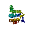
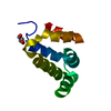
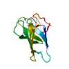
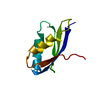
- PDB-1qys: Crystal structure of Top7: A computationally designed protein wit... -
+ Open data

- Basic information
| Entry | Database: PDB / ID: 1qys | ||||||
|---|---|---|---|---|---|---|---|
| Title | Crystal structure of Top7: A computationally designed protein with a novel fold | ||||||
 Components Components | TOP7 | ||||||
 Keywords Keywords | DE NOVO PROTEIN / alpha-beta / computationally designed / novel fold | ||||||
| Function / homology | top7, de novo designed protein / top7, de novo designed protein / 2-Layer Sandwich / Alpha Beta Function and homology information Function and homology information | ||||||
| Biological species | Computationally Designed Sequence | ||||||
| Method |  X-RAY DIFFRACTION / SYNCHROTRON / SAD / Resolution: 2.5 Å X-RAY DIFFRACTION / SYNCHROTRON / SAD / Resolution: 2.5 Å | ||||||
 Authors Authors | Kuhlman, B. / Dantas, G. / Ireton, G.C. / Varani, G. / Stoddard, B.L. / Baker, D. | ||||||
 Citation Citation | Journal: Science / Year: 2003 Title: Design of a Novel Globular Protein Fold with Atomic-Level Accuracy Authors: Kuhlman, B. / Dantas, G. / Ireton, G.C. / Varani, G. / Stoddard, B.L. / Baker, D. | ||||||
| History |
|
- Structure visualization
| Structure viewer | Molecule: MolmilJmol/JSmol |
|---|
- Downloads & links
-Download
| PDBx/mmCIF format | 1qys.cif.gz | 27.8 KB | Display | PDBx/mmCIF format |
|---|---|---|---|---|
| PDB format | pdb1qys.ent.gz | 17.9 KB | Display | PDB format |
| PDBx/mmJSON format | 1qys.json.gz | Tree view | PDBx/mmJSON format | |
| Others |  Other downloads Other downloads |
-Validation report
| Arichive directory | https://data.pdbj.org/pub/pdb/validation_reports/qy/1qysftp://data.pdbj.org/pub/pdb/validation_reports/qy/1qys | HTTPS FTP |
|---|
-Related structure data
| Similar structure data |
|---|


-Links
 PDBj
PDBj

- Assembly
| Deposited unit | 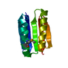
| ||||||||
|---|---|---|---|---|---|---|---|---|---|
| 1 |
| ||||||||
| Unit cell |
|
-Components
| #1: Protein | Mass: 12130.249 Da / Num. of mol.: 1 Source method: isolated from a genetically manipulated source Source: (gene. exp.) Computationally Designed Sequence / Plasmid: pet29b(+) / Production host:  |
|---|---|
| #2: Water | ChemComp-HOH /  Mass: 18.015 Da / Num. of mol.: 7 / Source method: isolated from a natural source / Formula: H2O Mass: 18.015 Da / Num. of mol.: 7 / Source method: isolated from a natural source / Formula: H2O |
| Has protein modification | Y |
-Experimental details
-Experiment
| Experiment | Method: X-RAY DIFFRACTION / Number of used crystals: 1 |
|---|
- Sample preparation
| Crystal | Density Matthews: 2.16 Å3/Da / Density % sol: 42.94 % | ||||||||||||||||||||||||
|---|---|---|---|---|---|---|---|---|---|---|---|---|---|---|---|---|---|---|---|---|---|---|---|---|---|
| Crystal grow | Temperature: 298 K / Method: vapor diffusion, hanging drop, streak seeding / pH: 6.6 Details: 15-20% PEG3350 250mM Ammonium Formate, pH 6.6, VAPOR DIFFUSION, HANGING DROP, STREAK SEEDING, temperature 298K | ||||||||||||||||||||||||
| Crystal grow | *PLUS Method: vapor diffusion, hanging drop | ||||||||||||||||||||||||
| Components of the solutions | *PLUS
|
-Data collection
| Diffraction | Mean temperature: 100 K |
|---|---|
| Diffraction source | Source: SYNCHROTRON / Site: ALS  / Beamline: 8.2.1 / Wavelength: 0.9793 Å / Beamline: 8.2.1 / Wavelength: 0.9793 Å |
| Detector | Type: ADSC QUANTUM 210 / Detector: CCD / Date: Mar 24, 2003 |
| Radiation | Monochromator: DOUBLE CRYSTAL Si(111) / Protocol: SAD / Monochromatic (M) / Laue (L): M / Scattering type: x-ray |
| Radiation wavelength | Wavelength: 0.9793 Å / Relative weight: 1 |
| Reflection | Resolution: 2.5→50 Å / Num. obs: 6979 / % possible obs: 100 % / Observed criterion σ(F): 0 / Observed criterion σ(I): 0 / Biso Wilson estimate: 30.2 Å2 / Rmerge(I) obs: 0.045 / Net I/σ(I): 37.8 |
| Reflection shell | Resolution: 2.5→2.59 Å / Rmerge(I) obs: 0.344 / Mean I/σ(I) obs: 5 / % possible all: 100 |
| Reflection | *PLUS Lowest resolution: 50 Å / Num. obs: 6989 / % possible obs: 99.1 % / Num. measured all: 144933 |
| Reflection shell | *PLUS % possible obs: 100 % |
- Processing
| Software |
| |||||||||||||||||||||||||
|---|---|---|---|---|---|---|---|---|---|---|---|---|---|---|---|---|---|---|---|---|---|---|---|---|---|---|
| Refinement | Method to determine structure: SAD / Resolution: 2.5→18.71 Å / Rfactor Rfree error: 0.016 / Data cutoff high absF: 1873860.4 / Data cutoff high rms absF: 1873860.4 / Data cutoff low absF: 0 / Isotropic thermal model: GROUP / Cross valid method: THROUGHOUT / σ(F): 0 / Stereochemistry target values: Engh & Huber
| |||||||||||||||||||||||||
| Solvent computation | Solvent model: FLAT MODEL / Bsol: 55.2431 Å2 / ksol: 0.300247 e/Å3 | |||||||||||||||||||||||||
| Displacement parameters | Biso mean: 65.5 Å2
| |||||||||||||||||||||||||
| Refine analyze |
| |||||||||||||||||||||||||
| Refinement step | Cycle: LAST / Resolution: 2.5→18.71 Å
| |||||||||||||||||||||||||
| Refine LS restraints |
| |||||||||||||||||||||||||
| LS refinement shell | Resolution: 2.5→2.66 Å / Rfactor Rfree error: 0.055 / Total num. of bins used: 6
| |||||||||||||||||||||||||
| Xplor file |
| |||||||||||||||||||||||||
| Refine LS restraints | *PLUS
|
