Protocol: SINGLE WAVELENGTH / Scattering type: x-ray
Radiation wavelength
Wavelength: 0.917 Å / Relative weight: 1
Reflection
Redundancy: 13.2 % / Av σ(I) over netI: 60.92 / Number: 365152 / Rmerge(I) obs: 0.13 / Χ2: 8.13 / D res high: 1.81 Å / D res low: 50 Å / Num. obs: 27686 / % possible obs: 98.3
Diffraction reflection shell
Highest resolution (Å)
Lowest resolution (Å)
% possible obs (%)
ID
Rmerge(I) obs
Chi squared
Redundancy
3.9
50
97
1
0.087
9.06
12.5
3.09
3.9
96.2
1
0.102
8.487
12.3
2.7
3.09
99.9
1
0.135
9.434
13.7
2.46
2.7
100
1
0.166
9.452
13.8
2.28
2.46
100
1
0.19
9.416
13.7
2.15
2.28
100
1
0.213
9.07
13.7
2.04
2.15
100
1
0.238
8.294
13.7
1.95
2.04
100
1
0.278
6.835
13.7
1.87
1.95
100
1
0.32
5.704
13.4
1.81
1.87
89.8
1
0.377
4.534
11.2
Reflection
Resolution: 1.81→50 Å / Num. obs: 27686 / % possible obs: 98.3 % / Redundancy: 13.2 % / Rmerge(I) obs: 0.13 / Χ2: 8.132 / Net I/σ(I): 14.5
Reflection shell
Resolution (Å)
Redundancy (%)
Rmerge(I) obs
Num. unique all
Χ2
Diffraction-ID
% possible all
1.81-1.87
11.2
0.377
2479
4.534
1
89.8
1.87-1.95
13.4
0.32
2803
5.704
1
100
1.95-2.04
13.7
0.278
2766
6.835
1
100
2.04-2.15
13.7
0.238
2777
8.294
1
100
2.15-2.28
13.7
0.213
2794
9.07
1
100
2.28-2.46
13.7
0.19
2802
9.416
1
100
2.46-2.7
13.8
0.166
2803
9.452
1
100
2.7-3.09
13.7
0.135
2831
9.434
1
99.9
3.09-3.9
12.3
0.102
2756
8.487
1
96.2
3.9-50
12.5
0.087
2875
9.06
1
97
-
Phasing
Phasing
Method: molecular replacement
-
Processing
Software
Name
Version
Classification
NB
DENZO
datareduction
SCALEPACK
datascaling
MOLREP
phasing
PHENIX
1.7.3_928
refinement
PDB_EXTRACT
3.11
dataextraction
Refinement
Method to determine structure: MOLECULAR REPLACEMENT / Resolution: 1.817→41.852 Å / Occupancy max: 1 / Occupancy min: 0.61 / FOM work R set: 0.7891 / SU ML: 0.25 / σ(F): 1.33 / Phase error: 26.87 / Stereochemistry target values: ML
Rfactor
Num. reflection
% reflection
Rfree
0.2421
1370
5.03 %
Rwork
0.2144
-
-
obs
0.2158
27254
97.02 %
Solvent computation
Shrinkage radii: 0.86 Å / VDW probe radii: 1.1 Å / Solvent model: FLAT BULK SOLVENT MODEL / Bsol: 42.389 Å2 / ksol: 0.404 e/Å3
In the structure databanks used in Yorodumi, some data are registered as the other names, "COVID-19 virus" and "2019-nCoV". Here are the details of the virus and the list of structure data.
Jan 31, 2019. EMDB accession codes are about to change! (news from PDBe EMDB page)
EMDB accession codes are about to change! (news from PDBe EMDB page)
The allocation of 4 digits for EMDB accession codes will soon come to an end. Whilst these codes will remain in use, new EMDB accession codes will include an additional digit and will expand incrementally as the available range of codes is exhausted. The current 4-digit format prefixed with “EMD-” (i.e. EMD-XXXX) will advance to a 5-digit format (i.e. EMD-XXXXX), and so on. It is currently estimated that the 4-digit codes will be depleted around Spring 2019, at which point the 5-digit format will come into force.
The EM Navigator/Yorodumi systems omit the EMD- prefix.
Related info.:Q: What is EMD? / ID/Accession-code notation in Yorodumi/EM Navigator
Yorodumi is a browser for structure data from EMDB, PDB, SASBDB, etc.
This page is also the successor to EM Navigator detail page, and also detail information page/front-end page for Omokage search.
The word "yorodu" (or yorozu) is an old Japanese word meaning "ten thousand". "mi" (miru) is to see.
Related info.:EMDB / PDB / SASBDB / Comparison of 3 databanks / Yorodumi Search / Aug 31, 2016. New EM Navigator & Yorodumi / Yorodumi Papers / Jmol/JSmol / Function and homology information / Changes in new EM Navigator and Yorodumi
 Movie
Movie Controller


 Open data
 Basic information
 Components
 Keywords
 Function and homology information
 Homo sapiens (human)
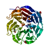
 X-RAY DIFFRACTION /
  Authors
 Citation
 Structure visualization
 Downloads & links
 Other downloads
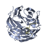


 PDBj
PDBj
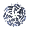
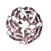
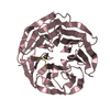
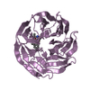
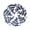
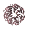
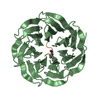
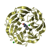
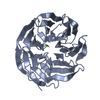
 Assembly
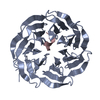

 Mass: 18.015 Da / Num. of mol.: 106 / Source method: isolated from a natural source / Formula: H2O
 Sample preparation
 / Beamline: F1 / Wavelength: 0.917 Å
 Processing