Protocol: SINGLE WAVELENGTH / Scattering type: x-ray
Radiation wavelength
Wavelength: 0.917 Å / Relative weight: 1
Reflection
Redundancy: 7.4 % / Av σ(I) over netI: 44.87 / Number: 719771 / Rmerge(I) obs: 0.079 / Χ2: 2.6 / D res high: 1.75 Å / D res low: 30 Å / Num. obs: 97077 / % possible obs: 99.8
Diffraction reflection shell
Highest resolution (Å)
Lowest resolution (Å)
% possible obs (%)
ID
Rmerge(I) obs
Chi squared
Redundancy
4.74
30
96.7
1
0.049
3.431
6.5
3.77
4.74
98.8
1
0.048
3.419
6.9
3.29
3.77
99.9
1
0.054
3.64
7.2
2.99
3.29
100
1
0.06
3.421
7.4
2.78
2.99
100
1
0.069
3.272
7.4
2.61
2.78
100
1
0.076
2.986
7.5
2.48
2.61
100
1
0.085
2.774
7.5
2.37
2.48
100
1
0.096
2.619
7.5
2.28
2.37
100
1
0.105
2.524
7.6
2.2
2.28
100
1
0.115
2.497
7.6
2.14
2.2
100
1
0.122
2.392
7.6
2.07
2.14
100
1
0.133
2.315
7.6
2.02
2.07
100
1
0.155
2.24
7.6
1.97
2.02
100
1
0.181
2.248
7.6
1.93
1.97
100
1
0.211
2.107
7.5
1.89
1.93
100
1
0.256
2.241
7.5
1.85
1.89
100
1
0.316
2.03
7.5
1.81
1.85
100
1
0.397
2.056
7.5
1.78
1.81
100
1
0.477
1.977
7.5
1.75
1.78
100
1
0.546
2.026
7.5
Reflection
Resolution: 1.75→30 Å / Num. obs: 97077 / % possible obs: 99.8 % / Redundancy: 7.4 % / Rmerge(I) obs: 0.079 / Net I/σ(I): 14.4
Reflection shell
Resolution (Å)
Redundancy (%)
Rmerge(I) obs
Diffraction-ID
% possible all
1.75-1.78
7.5
0.546
1
100
1.78-1.81
7.5
0.477
1
100
1.81-1.85
7.5
0.397
1
100
1.85-1.89
7.5
0.316
1
100
1.89-1.93
7.5
0.256
1
100
1.93-1.97
7.5
0.211
1
100
1.97-2.02
7.6
0.181
1
100
2.02-2.07
7.6
0.155
1
100
2.07-2.14
7.6
0.133
1
100
2.14-2.2
7.6
0.122
1
100
2.2-2.28
7.6
0.115
1
100
2.28-2.37
7.6
0.105
1
100
2.37-2.48
7.5
0.096
1
100
2.48-2.61
7.5
0.085
1
100
2.61-2.78
7.5
0.076
1
100
2.78-2.99
7.4
0.069
1
100
2.99-3.29
7.4
0.06
1
100
3.29-3.77
7.2
0.054
1
99.9
3.77-4.74
6.9
0.048
1
98.8
4.74-30
6.5
0.049
1
96.7
-
Phasing
Phasing
Method: molecular replacement
-
Processing
Software
Name
Version
Classification
NB
DENZO
datareduction
SCALEPACK
datascaling
MOLREP
phasing
PHENIX
1.7.3_928
refinement
PDB_EXTRACT
3.11
dataextraction
Refinement
Method to determine structure: MOLECULAR REPLACEMENT / Resolution: 1.75→27.24 Å / Occupancy max: 1 / Occupancy min: 0.32 / SU ML: 0.2 / σ(F): 1.33 / Phase error: 22.34 / Stereochemistry target values: ML
Rfactor
Num. reflection
% reflection
Rfree
0.216
4826
4.99 %
Rwork
0.186
-
-
obs
0.187
96631
99.5 %
Solvent computation
Shrinkage radii: 0.86 Å / VDW probe radii: 1.1 Å / Solvent model: FLAT BULK SOLVENT MODEL / Bsol: 40.45 Å2 / ksol: 0.4 e/Å3
Displacement parameters
Biso mean: 21.68 Å2
Baniso -1
Baniso -2
Baniso -3
1-
2.1791 Å2
0 Å2
-0.1456 Å2
2-
-
3.032 Å2
0 Å2
3-
-
-
-5.2111 Å2
Refinement step
Cycle: LAST / Resolution: 1.75→27.24 Å
Protein
Nucleic acid
Ligand
Solvent
Total
Num. atoms
7335
0
0
551
7886
Refine LS restraints
Refine-ID
Type
Dev ideal
Number
X-RAY DIFFRACTION
f_bond_d
0.007
7509
X-RAY DIFFRACTION
f_angle_d
1.174
10188
X-RAY DIFFRACTION
f_dihedral_angle_d
12.891
2652
X-RAY DIFFRACTION
f_chiral_restr
0.082
1146
X-RAY DIFFRACTION
f_plane_restr
0.006
1269
LS refinement shell
Resolution (Å)
Rfactor Rfree
Num. reflection Rfree
Rfactor Rwork
Num. reflection Rwork
Refine-ID
% reflection obs (%)
1.75-1.7699
0.2606
165
0.234
3001
X-RAY DIFFRACTION
99
1.7699-1.7907
0.3118
180
0.2397
3017
X-RAY DIFFRACTION
100
1.7907-1.8125
0.2482
174
0.235
3004
X-RAY DIFFRACTION
99
1.8125-1.8355
0.2798
136
0.2203
3088
X-RAY DIFFRACTION
99
1.8355-1.8596
0.2995
156
0.2209
3060
X-RAY DIFFRACTION
99
1.8596-1.8851
0.2491
146
0.2116
3060
X-RAY DIFFRACTION
100
1.8851-1.912
0.238
154
0.2068
3128
X-RAY DIFFRACTION
100
1.912-1.9405
0.2596
153
0.2028
2923
X-RAY DIFFRACTION
100
1.9405-1.9709
0.2305
176
0.1912
3044
X-RAY DIFFRACTION
100
1.9709-2.0032
0.2406
184
0.1887
3085
X-RAY DIFFRACTION
100
2.0032-2.0377
0.2315
136
0.1941
3069
X-RAY DIFFRACTION
100
2.0377-2.0747
0.2613
165
0.1968
3063
X-RAY DIFFRACTION
100
2.0747-2.1146
0.2446
142
0.1987
3120
X-RAY DIFFRACTION
100
2.1146-2.1578
0.2214
140
0.1912
3055
X-RAY DIFFRACTION
100
2.1578-2.2047
0.2517
165
0.1859
3052
X-RAY DIFFRACTION
100
2.2047-2.2559
0.2316
170
0.1804
3077
X-RAY DIFFRACTION
100
2.2559-2.3123
0.2188
179
0.191
3032
X-RAY DIFFRACTION
100
2.3123-2.3748
0.2398
162
0.191
3097
X-RAY DIFFRACTION
100
2.3748-2.4446
0.2004
158
0.1937
3057
X-RAY DIFFRACTION
100
2.4446-2.5235
0.2502
172
0.2056
3077
X-RAY DIFFRACTION
100
2.5235-2.6136
0.224
139
0.1989
3057
X-RAY DIFFRACTION
100
2.6136-2.7182
0.2419
162
0.2011
3034
X-RAY DIFFRACTION
100
2.7182-2.8418
0.2393
181
0.2088
3101
X-RAY DIFFRACTION
100
2.8418-2.9914
0.222
165
0.2029
3078
X-RAY DIFFRACTION
100
2.9914-3.1786
0.2193
152
0.1946
3056
X-RAY DIFFRACTION
100
3.1786-3.4235
0.1921
144
0.1737
3112
X-RAY DIFFRACTION
100
3.4235-3.7673
0.1876
171
0.1642
3087
X-RAY DIFFRACTION
100
3.7673-4.3105
0.1663
162
0.1537
3056
X-RAY DIFFRACTION
99
4.3105-5.4238
0.16
182
0.1449
3044
X-RAY DIFFRACTION
98
5.4238-27.239
0.2208
155
0.1882
3071
X-RAY DIFFRACTION
96
+
About Yorodumi
-
News
-
Feb 9, 2022. New format data for meta-information of EMDB entries
New format data for meta-information of EMDB entries
Version 3 of the EMDB header file is now the official format.
The previous official version 1.9 will be removed from the archive.
In the structure databanks used in Yorodumi, some data are registered as the other names, "COVID-19 virus" and "2019-nCoV". Here are the details of the virus and the list of structure data.
Jan 31, 2019. EMDB accession codes are about to change! (news from PDBe EMDB page)
EMDB accession codes are about to change! (news from PDBe EMDB page)
The allocation of 4 digits for EMDB accession codes will soon come to an end. Whilst these codes will remain in use, new EMDB accession codes will include an additional digit and will expand incrementally as the available range of codes is exhausted. The current 4-digit format prefixed with “EMD-” (i.e. EMD-XXXX) will advance to a 5-digit format (i.e. EMD-XXXXX), and so on. It is currently estimated that the 4-digit codes will be depleted around Spring 2019, at which point the 5-digit format will come into force.
The EM Navigator/Yorodumi systems omit the EMD- prefix.
Related info.:Q: What is EMD? / ID/Accession-code notation in Yorodumi/EM Navigator
Yorodumi is a browser for structure data from EMDB, PDB, SASBDB, etc.
This page is also the successor to EM Navigator detail page, and also detail information page/front-end page for Omokage search.
The word "yorodu" (or yorozu) is an old Japanese word meaning "ten thousand". "mi" (miru) is to see.
Related info.:EMDB / PDB / SASBDB / Comparison of 3 databanks / Yorodumi Search / Aug 31, 2016. New EM Navigator & Yorodumi / Yorodumi Papers / Jmol/JSmol / Function and homology information / Changes in new EM Navigator and Yorodumi
 Movie
Movie Controller


 Open data
 Basic information
 Components
 Keywords
 Function and homology information
 Homo sapiens (human)
 X-RAY DIFFRACTION /
  Authors
 Citation
 Structure visualization
 Downloads & links
 Other downloads
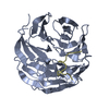


 PDBj
PDBj

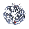
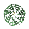
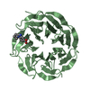
 Assembly


 Mass: 18.015 Da / Num. of mol.: 551 / Source method: isolated from a natural source / Formula: H2O
 Sample preparation
 / Beamline: F1 / Wavelength: 0.917 Å
 Processing