 Movie
Movie Controller


+ Open data

- Basic information
| Entry | Database: PDB / ID: 2xl3 | ||||||
|---|---|---|---|---|---|---|---|
| Title | WDR5 IN COMPLEX WITH AN RBBP5 PEPTIDE AND HISTONE H3 PEPTIDE | ||||||
 Components Components |
| ||||||
 Keywords Keywords | TRANSCRIPTION / MLL COMPLEX / H3K4 METHYLATION / WD-40 BETA-PROPELLER | ||||||
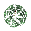
| Function / homology |  Function and homology information Function and homology informationEpigenetic regulation of gene expression by MLL3 and MLL4 complexes / Formation of WDR5-containing histone-modifying complexes / Chromatin modifying enzymes / Interleukin-7 signaling / Regulation of endogenous retroelements by KRAB-ZFP proteins / Condensation of Prophase Chromosomes / HDMs demethylate histones / Negative Regulation of CDH1 Gene Transcription / HDACs deacetylate histones / Formation of the beta-catenin:TCF transactivating complex ...Epigenetic regulation of gene expression by MLL3 and MLL4 complexes / Formation of WDR5-containing histone-modifying complexes / Chromatin modifying enzymes / Interleukin-7 signaling / Regulation of endogenous retroelements by KRAB-ZFP proteins / Condensation of Prophase Chromosomes / HDMs demethylate histones / Negative Regulation of CDH1 Gene Transcription / HDACs deacetylate histones / Formation of the beta-catenin:TCF transactivating complex / PRC2 methylates histones and DNA / HATs acetylate histones / MLL4 and MLL3 complexes regulate expression of PPARG target genes in adipogenesis and hepatic steatosis / PKMTs methylate histone lysines / RUNX1 regulates genes involved in megakaryocyte differentiation and platelet function / RMTs methylate histone arginines / Factors involved in megakaryocyte development and platelet production / Estrogen-dependent gene expression / histone H3Q5ser reader activity / Neddylation / histone H3K4me1 reader activity / MLL3/4 complex / Set1C/COMPASS complex / ATAC complex / MLL1/2 complex / NSL complex / histone H3K4 methyltransferase activity / histone methyltransferase complex / MLL1 complex / histone acetyltransferase complex / regulation of embryonic development / regulation of cell division / positive regulation of gluconeogenesis / transcription initiation-coupled chromatin remodeling / skeletal system development / response to estrogen / neuron projection development / structural constituent of chromatin / mitotic spindle / nucleosome / chromatin organization / histone binding / gene expression / regulation of cell cycle / transcription cis-regulatory region binding / protein heterodimerization activity / DNA damage response / regulation of transcription by RNA polymerase II / regulation of DNA-templated transcription / positive regulation of DNA-templated transcription / nucleolus / negative regulation of transcription by RNA polymerase II / protein-containing complex / DNA binding / nucleoplasm / nucleus Similarity search - Function | ||||||
| Biological species |  | ||||||
| Method |  X-RAY DIFFRACTION / SYNCHROTRON / MOLECULAR REPLACEMENT / Resolution: 2.7 Å X-RAY DIFFRACTION / SYNCHROTRON / MOLECULAR REPLACEMENT / Resolution: 2.7 Å | ||||||
 Authors Authors | Odho, Z. / Southall, S.M. / Wilson, J.R. | ||||||
 Citation Citation | Journal: J.Biol.Chem. / Year: 2010 Title: Characterisation of a Novel Wdr5 Binding Site that Recruits Rbbp5 Through a Conserved Motif and Enhances Methylation of H3K4 by Mll1. Authors: Odho, Z. / Southall, S.M. / Wilson, J.R. | ||||||
| History |
|
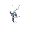
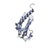
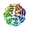
- Structure visualization
| Structure viewer | Molecule: MolmilJmol/JSmol |
|---|
- Downloads & links
-Download
| PDBx/mmCIF format | 2xl3.cif.gz | 135.6 KB | Display | PDBx/mmCIF format |
|---|---|---|---|---|
| PDB format | pdb2xl3.ent.gz | 105.1 KB | Display | PDB format |
| PDBx/mmJSON format | 2xl3.json.gz | Tree view | PDBx/mmJSON format | |
| Others |  Other downloads Other downloads |
-Validation report
| Arichive directory | https://data.pdbj.org/pub/pdb/validation_reports/xl/2xl3ftp://data.pdbj.org/pub/pdb/validation_reports/xl/2xl3 | HTTPS FTP |
|---|
-Related structure data
| Related structure data | 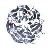 2xl2C  2h13S C: citing same article ( S: Starting model for refinement |
|---|---|
| Similar structure data |


-Links
 PDBj
PDBj


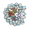
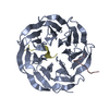
- Assembly
| Deposited unit | 
| ||||||||||||||||||
|---|---|---|---|---|---|---|---|---|---|---|---|---|---|---|---|---|---|---|---|
| 1 | 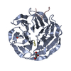
| ||||||||||||||||||
| 2 | 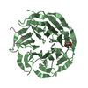
| ||||||||||||||||||
| Unit cell |
| ||||||||||||||||||
| Noncrystallographic symmetry (NCS) | NCS domain:
NCS domain segments:
NCS oper: (Code: given Matrix: (0.998316, -0.000389, 0.058006), Vector: |
-Components
| #1: Protein | Mass: 36635.438 Da / Num. of mol.: 2 Source method: isolated from a genetically manipulated source Source: (gene. exp.)  #2: Protein/peptide | Mass: 1541.524 Da / Num. of mol.: 2 / Fragment: RESIDUES 369-381 / Source method: obtained synthetically / Source: (synth.) #3: Protein/peptide | Mass: 1097.248 Da / Num. of mol.: 2 / Fragment: AMINO TERMINAL TAIL, RESIDUES 2-9 / Source method: obtained synthetically / Source: (synth.) #4: Chemical |   Mass: 92.094 Da / Num. of mol.: 3 / Source method: obtained synthetically / Formula: C3H8O3 Mass: 92.094 Da / Num. of mol.: 3 / Source method: obtained synthetically / Formula: C3H8O3#5: Water | ChemComp-HOH / |  Mass: 18.015 Da / Num. of mol.: 107 / Source method: isolated from a natural source / Formula: H2O Mass: 18.015 Da / Num. of mol.: 107 / Source method: isolated from a natural source / Formula: H2OSequence details | NON NATIVE CARBOXY TERMINAL TYROSINE NON NATIVE TYROSINE AT AMINO TERMINAL | |
|---|
-Experimental details
-Experiment
| Experiment | Method: X-RAY DIFFRACTION / Number of used crystals: 1 |
|---|
- Sample preparation
| Crystal | Density Matthews: 2.45 Å3/Da / Density % sol: 49.7 % / Description: NONE |
|---|---|
| Crystal grow | pH: 8 / Details: 0.2M K2SO4 20 % PEG 3350, pH 8.0 |
-Data collection
| Diffraction | Mean temperature: 80 K |
|---|---|
| Diffraction source | Source: SYNCHROTRON / Site: ESRF  / Beamline: ID14-2 / Wavelength: 0.933 / Beamline: ID14-2 / Wavelength: 0.933 |
| Detector | Type: ADSC CCD / Detector: CCD / Date: Nov 25, 2009 / Details: MIRRORS |
| Radiation | Protocol: SINGLE WAVELENGTH / Monochromatic (M) / Laue (L): M / Scattering type: x-ray |
| Radiation wavelength | Wavelength: 0.933 Å / Relative weight: 1 |
| Reflection | Resolution: 2.7→86.3 Å / Num. obs: 16657 / % possible obs: 98.7 % / Observed criterion σ(I): 2 / Redundancy: 3 % / Biso Wilson estimate: 32 Å2 / Rmerge(I) obs: 0.11 / Net I/σ(I): 7.2 |
| Reflection shell | Resolution: 2.7→2.9 Å / Redundancy: 3 % / Rmerge(I) obs: 0.46 / Mean I/σ(I) obs: 2.5 / % possible all: 99 |
- Processing
| Software |
| |||||||||||||||||||||||||||||||||||||||||||||||||
|---|---|---|---|---|---|---|---|---|---|---|---|---|---|---|---|---|---|---|---|---|---|---|---|---|---|---|---|---|---|---|---|---|---|---|---|---|---|---|---|---|---|---|---|---|---|---|---|---|---|---|
| Refinement | Method to determine structure: MOLECULAR REPLACEMENT Starting model: PDB ENTRY 2H13 Resolution: 2.7→41.688 Å / SU ML: 0.3 / σ(F): 0.02 / Phase error: 27.11 / Stereochemistry target values: ML
| |||||||||||||||||||||||||||||||||||||||||||||||||
| Solvent computation | Shrinkage radii: 0.9 Å / VDW probe radii: 1.11 Å / Solvent model: FLAT BULK SOLVENT MODEL / Bsol: 25.69 Å2 / ksol: 0.323 e/Å3 | |||||||||||||||||||||||||||||||||||||||||||||||||
| Displacement parameters | Biso mean: 37.1 Å2
| |||||||||||||||||||||||||||||||||||||||||||||||||
| Refinement step | Cycle: LAST / Resolution: 2.7→41.688 Å
| |||||||||||||||||||||||||||||||||||||||||||||||||
| Refine LS restraints |
| |||||||||||||||||||||||||||||||||||||||||||||||||
| Refine LS restraints NCS |
| |||||||||||||||||||||||||||||||||||||||||||||||||
| LS refinement shell |
|
