Mass: 18.015 Da / Num. of mol.: 211 / Source method: isolated from a natural source / Formula: H2O
Has protein modification
Y
-
Experimental details
-
Experiment
Experiment
Method: X-RAY DIFFRACTION / Number of used crystals: 1
-
Sample preparation
Crystal
Density Matthews: 3.23 Å3/Da / Density % sol: 62 % / Description: NONE
Crystal grow
Method: vapor diffusion, hanging drop / pH: 7.5 Details: BRDT BD2 PROTEIN AT 25 MG/ML WAS MIXED WITH H3-ACK18 PEPTIDE IN A 1:5 MOLAR RATIO. CRYSTALLIZATION WAS BY THE HANGING DROP VAPOUR DIFFUSION METHOD USING 2.0 M AMMONIUM SULFATE, 2% PEG 400, 0.1 M HEPES PH 7.5
Resolution: 2.1→40 Å / Rfactor Rfree error: 0.008 / Data cutoff high absF: 100000000 / Data cutoff low absF: 0 / Cross valid method: THROUGHOUT / σ(F): 0 / Stereochemistry target values: MAXIMUM LIKELIHOOD Details: THE FOLLOWING RESIDUES WERE NOT MODELLED DUE TO POOR DENSITY - CHAIN A RESIDUES 257-258 AND 382, CHAIN B RESIDUES 257-264 AND 377-382, CHAIN P RESIDUES 14 AND 22-23. THE FOLLOWING RESIDUES ...Details: THE FOLLOWING RESIDUES WERE NOT MODELLED DUE TO POOR DENSITY - CHAIN A RESIDUES 257-258 AND 382, CHAIN B RESIDUES 257-264 AND 377-382, CHAIN P RESIDUES 14 AND 22-23. THE FOLLOWING RESIDUES WERE MODELLED AS GLYCINE -CHAIN A RESIDUES 260-261 AND 381, CHAIN B RESIDUES 265-267, CHAIN Q RESIDUE 14.
In the structure databanks used in Yorodumi, some data are registered as the other names, "COVID-19 virus" and "2019-nCoV". Here are the details of the virus and the list of structure data.
Jan 31, 2019. EMDB accession codes are about to change! (news from PDBe EMDB page)
EMDB accession codes are about to change! (news from PDBe EMDB page)
The allocation of 4 digits for EMDB accession codes will soon come to an end. Whilst these codes will remain in use, new EMDB accession codes will include an additional digit and will expand incrementally as the available range of codes is exhausted. The current 4-digit format prefixed with “EMD-” (i.e. EMD-XXXX) will advance to a 5-digit format (i.e. EMD-XXXXX), and so on. It is currently estimated that the 4-digit codes will be depleted around Spring 2019, at which point the 5-digit format will come into force.
The EM Navigator/Yorodumi systems omit the EMD- prefix.
Related info.:Q: What is EMD? / ID/Accession-code notation in Yorodumi/EM Navigator
Yorodumi is a browser for structure data from EMDB, PDB, SASBDB, etc.
This page is also the successor to EM Navigator detail page, and also detail information page/front-end page for Omokage search.
The word "yorodu" (or yorozu) is an old Japanese word meaning "ten thousand". "mi" (miru) is to see.
Related info.:EMDB / PDB / SASBDB / Comparison of 3 databanks / Yorodumi Search / Aug 31, 2016. New EM Navigator & Yorodumi / Yorodumi Papers / Jmol/JSmol / Function and homology information / Changes in new EM Navigator and Yorodumi
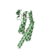
 Movie
Movie Controller

 Yorodumi
 Open data
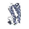
 Basic information
 Components
 Keywords
 Function and homology information
 X-RAY DIFFRACTION /
  Authors
 Citation
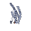
 Structure visualization
 Downloads & links
 Other downloads
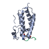


 PDBj
PDBj


 Assembly


 Mass: 18.015 Da / Num. of mol.: 211 / Source method: isolated from a natural source / Formula: H2O
 Sample preparation
 / Beamline: ID23-1 / Wavelength: 1.07225
  Processing