 Movie
Movie Controller


+ Open data

- Basic information
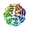
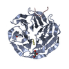
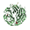
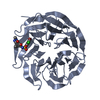
| Entry | Database: PDB / ID: 4cy2 | ||||||
|---|---|---|---|---|---|---|---|
| Title | Crystal structure of the KANSL1-WDR5-KANSL2 complex. | ||||||
 Components Components |
| ||||||
 Keywords Keywords | TRANSCRIPTION / EPIGENETIC REGULATOR / HISTONE ACETYLATION / CHROMATIN | ||||||
| Function / homology |  Function and homology information Function and homology informationregulation of mitochondrial transcription / histone H3Q5ser reader activity / histone H3K4me1 reader activity / Loss of Function of KMT2D in MLL4 Complex Formation in Kabuki Syndrome / Epigenetic regulation of gene expression by MLL3 and MLL4 complexes / MLL3/4 complex / Set1C/COMPASS complex / ATAC complex / MLL1/2 complex / NSL complex ...regulation of mitochondrial transcription / histone H3Q5ser reader activity / histone H3K4me1 reader activity / Loss of Function of KMT2D in MLL4 Complex Formation in Kabuki Syndrome / Epigenetic regulation of gene expression by MLL3 and MLL4 complexes / MLL3/4 complex / Set1C/COMPASS complex / ATAC complex / MLL1/2 complex / NSL complex / histone H3K4 methyltransferase activity / Cardiogenesis / histone acetyltransferase binding / Formation of WDR5-containing histone-modifying complexes / histone methyltransferase complex / MLL1 complex / histone acetyltransferase complex / regulation of embryonic development / regulation of cell division / positive regulation of gluconeogenesis / transcription initiation-coupled chromatin remodeling / gluconeogenesis / skeletal system development / RUNX1 regulates genes involved in megakaryocyte differentiation and platelet function / kinetochore / PKMTs methylate histone lysines / Activation of anterior HOX genes in hindbrain development during early embryogenesis / RMTs methylate histone arginines / spindle pole / mitotic spindle / actin cytoskeleton / HATs acetylate histones / MLL4 and MLL3 complexes regulate expression of PPARG target genes in adipogenesis and hepatic steatosis / sperm principal piece / chromatin organization / Neddylation / histone binding / microtubule / regulation of cell cycle / regulation of transcription by RNA polymerase II / regulation of DNA-templated transcription / positive regulation of DNA-templated transcription / negative regulation of transcription by RNA polymerase II / mitochondrion / nucleoplasm / nucleus / plasma membrane / cytosol Similarity search - Function | ||||||
| Biological species |  HOMO SAPIENS (human) HOMO SAPIENS (human) | ||||||
| Method |  X-RAY DIFFRACTION / SYNCHROTRON / MOLECULAR REPLACEMENT / Resolution: 2 Å X-RAY DIFFRACTION / SYNCHROTRON / MOLECULAR REPLACEMENT / Resolution: 2 Å | ||||||
 Authors Authors | Dias, J. / Brettschneider, J. / Cusack, S. / Kadlec, J. | ||||||
 Citation Citation | Journal: Genes Dev. / Year: 2014 Title: Structural Analysis of the Kansl1/Wdr5/Kansl2 Complex Reveals that Wdr5 is Required for Efficient Assembly and Chromatin Targeting of the Nsl Complex. Authors: Dias, J. / Van Nguyen, N. / Georgiev, P. / Gaub, A. / Brettschneider, J. / Cusack, S. / Kadlec, J. / Akhtar, A. | ||||||
| History |
|
- Structure visualization
| Structure viewer | Molecule: MolmilJmol/JSmol |
|---|
- Downloads & links
-Download
| PDBx/mmCIF format | 4cy2.cif.gz | 138 KB | Display | PDBx/mmCIF format |
|---|---|---|---|---|
| PDB format | pdb4cy2.ent.gz | 108 KB | Display | PDB format |
| PDBx/mmJSON format | 4cy2.json.gz | Tree view | PDBx/mmJSON format | |
| Others |  Other downloads Other downloads |
-Validation report
| Arichive directory | https://data.pdbj.org/pub/pdb/validation_reports/cy/4cy2ftp://data.pdbj.org/pub/pdb/validation_reports/cy/4cy2 | HTTPS FTP |
|---|
-Related structure data
| Related structure data |  4cy1C  4cy3C  4cy5C  2g99S C: citing same article ( S: Starting model for refinement |
|---|---|
| Similar structure data |


-Links
 PDBj
PDBj
- Assembly
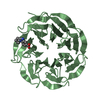
| Deposited unit | 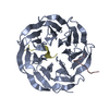
| ||||||||
|---|---|---|---|---|---|---|---|---|---|
| 1 |
| ||||||||
| Unit cell |
|
-Components
| #1: Protein | Mass: 34620.289 Da / Num. of mol.: 1 / Fragment: RESIDUES 23-334 Source method: isolated from a genetically manipulated source Source: (gene. exp.) HOMO SAPIENS (human) / Plasmid: PPROEXHTB / Production host:  |
|---|---|
| #2: Protein/peptide | Mass: 1430.429 Da / Num. of mol.: 1 / Fragment: RESIDUES 406-417 / Source method: obtained synthetically / Source: (synth.) HOMO SAPIENS (human) / References: UniProt: Q9H9L4 |
| #3: Protein/peptide | Mass: 1610.834 Da / Num. of mol.: 1 / Fragment: RESIDUES 585-598 / Source method: obtained synthetically / Source: (synth.) HOMO SAPIENS (human) / References: UniProt: Q7Z3B3 |
| #4: Water | ChemComp-HOH /  Mass: 18.015 Da / Num. of mol.: 104 / Source method: isolated from a natural source / Formula: H2O Mass: 18.015 Da / Num. of mol.: 104 / Source method: isolated from a natural source / Formula: H2O |
-Experimental details
-Experiment
| Experiment | Method: X-RAY DIFFRACTION / Number of used crystals: 1 |
|---|
- Sample preparation
| Crystal | Density Matthews: 2.14 Å3/Da / Density % sol: 0.43 % / Description: NONE |
|---|---|
| Crystal grow | pH: 6.9 Details: 20% (W/V) PEG 3350, 0.2 M POTASSIUM NITRATE, PH 6.9. |
-Data collection
| Diffraction | Mean temperature: 100 K |
|---|---|
| Diffraction source | Source: SYNCHROTRON / Site: ESRF  / Beamline: ID23-2 / Wavelength: 0.8726 / Beamline: ID23-2 / Wavelength: 0.8726 |
| Detector | Type: MARRESEARCH / Detector: CCD / Date: May 2, 2013 |
| Radiation | Protocol: SINGLE WAVELENGTH / Monochromatic (M) / Laue (L): M / Scattering type: x-ray |
| Radiation wavelength | Wavelength: 0.8726 Å / Relative weight: 1 |
| Reflection | Resolution: 2→100 Å / Num. obs: 22261 / % possible obs: 100 % / Observed criterion σ(I): 2.6 / Redundancy: 7.3 % / Rmerge(I) obs: 0.15 / Net I/σ(I): 10.7 |
| Reflection shell | Resolution: 2→2.1 Å / Redundancy: 7.4 % / Rmerge(I) obs: 0.84 / Mean I/σ(I) obs: 2.6 / % possible all: 99.9 |
- Processing
| Software |
| ||||||||||||||||||||||||||||||||||||||||||||||||||||||||||||||||||||||||||||||||||||||||||||||||||||||||||||||||||||||||||||||||||||||||||||||||||||||||||||||||||||||||||||||||||||||
|---|---|---|---|---|---|---|---|---|---|---|---|---|---|---|---|---|---|---|---|---|---|---|---|---|---|---|---|---|---|---|---|---|---|---|---|---|---|---|---|---|---|---|---|---|---|---|---|---|---|---|---|---|---|---|---|---|---|---|---|---|---|---|---|---|---|---|---|---|---|---|---|---|---|---|---|---|---|---|---|---|---|---|---|---|---|---|---|---|---|---|---|---|---|---|---|---|---|---|---|---|---|---|---|---|---|---|---|---|---|---|---|---|---|---|---|---|---|---|---|---|---|---|---|---|---|---|---|---|---|---|---|---|---|---|---|---|---|---|---|---|---|---|---|---|---|---|---|---|---|---|---|---|---|---|---|---|---|---|---|---|---|---|---|---|---|---|---|---|---|---|---|---|---|---|---|---|---|---|---|---|---|---|---|
| Refinement | Method to determine structure: MOLECULAR REPLACEMENT Starting model: PDB ENTRY 2G99 Resolution: 2→43.3 Å / Cor.coef. Fo:Fc: 0.947 / Cor.coef. Fo:Fc free: 0.946 / SU B: 5.415 / SU ML: 0.08 / Cross valid method: THROUGHOUT / ESU R: 0.043 / ESU R Free: 0.034 / Stereochemistry target values: MAXIMUM LIKELIHOOD Details: HYDROGENS HAVE BEEN ADDED IN THE RIDING POSITIONS. U VALUES WITH TLS ADDED
| ||||||||||||||||||||||||||||||||||||||||||||||||||||||||||||||||||||||||||||||||||||||||||||||||||||||||||||||||||||||||||||||||||||||||||||||||||||||||||||||||||||||||||||||||||||||
| Solvent computation | Ion probe radii: 0.8 Å / Shrinkage radii: 0.8 Å / VDW probe radii: 1.2 Å / Solvent model: MASK | ||||||||||||||||||||||||||||||||||||||||||||||||||||||||||||||||||||||||||||||||||||||||||||||||||||||||||||||||||||||||||||||||||||||||||||||||||||||||||||||||||||||||||||||||||||||
| Displacement parameters | Biso mean: 24.616 Å2
| ||||||||||||||||||||||||||||||||||||||||||||||||||||||||||||||||||||||||||||||||||||||||||||||||||||||||||||||||||||||||||||||||||||||||||||||||||||||||||||||||||||||||||||||||||||||
| Refinement step | Cycle: LAST / Resolution: 2→43.3 Å
| ||||||||||||||||||||||||||||||||||||||||||||||||||||||||||||||||||||||||||||||||||||||||||||||||||||||||||||||||||||||||||||||||||||||||||||||||||||||||||||||||||||||||||||||||||||||
| Refine LS restraints |
|