 Movie
Movie Controller

[English] 日本語
 Yorodumi
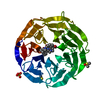
- PDB-2g99: Structural basis for the specific recognition of methylated histo... -
+ Open data

- Basic information
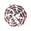
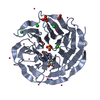
| Entry | Database: PDB / ID: 2g99 | ||||||
|---|---|---|---|---|---|---|---|
| Title | Structural basis for the specific recognition of methylated histone H3 lysine 4 by the WD-40 protein WDR5 | ||||||
 Components Components |
| ||||||
 Keywords Keywords | STRUCTURAL PROTEIN/DNA BINDING PROTEIN / WD40 repeat protein / STRUCTURAL PROTEIN-DNA BINDING PROTEIN COMPLEX | ||||||
| Function / homology |  Function and homology information Function and homology informationhistone H3Q5ser reader activity / histone H3K4me1 reader activity / Loss of Function of KMT2D in MLL4 Complex Formation in Kabuki Syndrome / Epigenetic regulation of gene expression by MLL3 and MLL4 complexes / MLL3/4 complex / Set1C/COMPASS complex / ATAC complex / MLL1/2 complex / NSL complex / histone H3K4 methyltransferase activity ...histone H3Q5ser reader activity / histone H3K4me1 reader activity / Loss of Function of KMT2D in MLL4 Complex Formation in Kabuki Syndrome / Epigenetic regulation of gene expression by MLL3 and MLL4 complexes / MLL3/4 complex / Set1C/COMPASS complex / ATAC complex / MLL1/2 complex / NSL complex / histone H3K4 methyltransferase activity / Cardiogenesis / Formation of WDR5-containing histone-modifying complexes / histone methyltransferase complex / MLL1 complex / histone acetyltransferase complex / regulation of embryonic development / regulation of cell division / positive regulation of gluconeogenesis / transcription initiation-coupled chromatin remodeling / gluconeogenesis / skeletal system development / RUNX1 regulates genes involved in megakaryocyte differentiation and platelet function / PKMTs methylate histone lysines / Activation of anterior HOX genes in hindbrain development during early embryogenesis / RMTs methylate histone arginines / mitotic spindle / HATs acetylate histones / MLL4 and MLL3 complexes regulate expression of PPARG target genes in adipogenesis and hepatic steatosis / sperm principal piece / Neddylation / histone binding / regulation of cell cycle / regulation of transcription by RNA polymerase II / regulation of DNA-templated transcription / positive regulation of DNA-templated transcription / negative regulation of transcription by RNA polymerase II / nucleoplasm / nucleus Similarity search - Function | ||||||
| Biological species |  Homo sapiens (human) Homo sapiens (human) | ||||||
| Method |  X-RAY DIFFRACTION / MOLECULAR REPLACEMENT / Resolution: 1.9 Å X-RAY DIFFRACTION / MOLECULAR REPLACEMENT / Resolution: 1.9 Å | ||||||
 Authors Authors | Chai, J. / Han, Z. / Wang, H. / Shen, Y. | ||||||
 Citation Citation | Journal: To be published Title: Structural basis for the specific recognition of methylated histone H3 lysine 4 by the WD-40 protein WDR5 Authors: Chai, J. / Han, Z. / Wang, H. / Shen, Y. | ||||||
| History |
|
- Structure visualization
| Structure viewer | Molecule: MolmilJmol/JSmol |
|---|
- Downloads & links
-Download
| PDBx/mmCIF format | 2g99.cif.gz | 126.7 KB | Display | PDBx/mmCIF format |
|---|---|---|---|---|
| PDB format | pdb2g99.ent.gz | 98.3 KB | Display | PDB format |
| PDBx/mmJSON format | 2g99.json.gz | Tree view | PDBx/mmJSON format | |
| Others |  Other downloads Other downloads |
-Validation report
| Arichive directory | https://data.pdbj.org/pub/pdb/validation_reports/g9/2g99ftp://data.pdbj.org/pub/pdb/validation_reports/g9/2g99 | HTTPS FTP |
|---|
-Related structure data
| Related structure data |  2g9aC  1vyhS C: citing same article ( S: Starting model for refinement |
|---|---|
| Similar structure data |


-Links
 PDBj
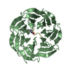
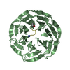
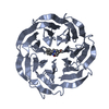
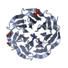
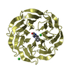
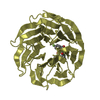
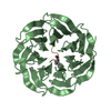
PDBj- Assembly
| Deposited unit | 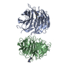
| ||||||||
|---|---|---|---|---|---|---|---|---|---|
| 1 | 
| ||||||||
| 2 | 
| ||||||||
| Unit cell |
|
-Components
| #1: Protein | Mass: 33916.527 Da / Num. of mol.: 2 / Fragment: residues 27-334 Source method: isolated from a genetically manipulated source Source: (gene. exp.) Homo sapiens (human) / Production host:  #2: Protein/peptide | Mass: 1204.424 Da / Num. of mol.: 2 / Source method: obtained synthetically Details: The dimethylated tail of histone H3 is chemically synthesized |
|---|
-Experimental details
-Experiment
| Experiment | Method: X-RAY DIFFRACTION / Number of used crystals: 1 |
|---|
- Sample preparation
| Crystal | Density Matthews: 2.23 Å3/Da / Density % sol: 44.9 % |
|---|---|
| Crystal grow | Temperature: 298 K / Method: vapor diffusion, hanging drop / pH: 7.5 Details: 20% PEG 4000, 10% isopropanol, 0.1M HEPES, pH7.5 , VAPOR DIFFUSION, HANGING DROP, temperature 298.0K |
-Data collection
| Diffraction | Mean temperature: 180 K |
|---|---|
| Diffraction source | Source: ROTATING ANODE / Type: RIGAKU RU300 / Wavelength: 1.5418 Å |
| Detector | Type: RIGAKU RAXIS IV / Detector: IMAGE PLATE / Date: Nov 10, 2005 / Details: mirrors |
| Radiation | Monochromator: YALE MIRRORS / Protocol: SINGLE WAVELENGTH / Monochromatic (M) / Laue (L): M / Scattering type: x-ray |
| Radiation wavelength | Wavelength: 1.5418 Å / Relative weight: 1 |
| Reflection | Resolution: 1.9→99 Å / Num. all: 49725 / Num. obs: 45846 / % possible obs: 92.2 % / Observed criterion σ(F): 2 / Observed criterion σ(I): 1 |
| Reflection shell | Resolution: 1.9→1.97 Å / % possible all: 87.4 |
- Processing
| Software |
| ||||||||||||||||||||
|---|---|---|---|---|---|---|---|---|---|---|---|---|---|---|---|---|---|---|---|---|---|
| Refinement | Method to determine structure: MOLECULAR REPLACEMENT Starting model: PDB ENTRY 1VYH Resolution: 1.9→20 Å / σ(F): 0 / Stereochemistry target values: Engh & Huber
| ||||||||||||||||||||
| Refinement step | Cycle: LAST / Resolution: 1.9→20 Å
| ||||||||||||||||||||
| Refine LS restraints |
|
