 Movie
Movie Controller


+ Open data

- Basic information
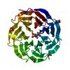
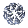
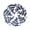
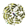
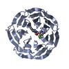
| Entry | Database: PDB / ID: 6oi0 | ||||||
|---|---|---|---|---|---|---|---|
| Title | Crystal structure of human WDR5 in complex with L-arginine | ||||||
 Components Components | WD repeat-containing protein 5 | ||||||
 Keywords Keywords | GENE REGULATION / Methylation / Epigenetics / Post-translational modification / Chromatin / Methylarginine | ||||||
| Function / homology |  Function and homology information Function and homology informationhistone H3Q5ser reader activity / histone H3K4me1 reader activity / Loss of Function of KMT2D in MLL4 Complex Formation in Kabuki Syndrome / Epigenetic regulation of gene expression by MLL3 and MLL4 complexes / MLL3/4 complex / Set1C/COMPASS complex / ATAC complex / MLL1/2 complex / NSL complex / histone H3K4 methyltransferase activity ...histone H3Q5ser reader activity / histone H3K4me1 reader activity / Loss of Function of KMT2D in MLL4 Complex Formation in Kabuki Syndrome / Epigenetic regulation of gene expression by MLL3 and MLL4 complexes / MLL3/4 complex / Set1C/COMPASS complex / ATAC complex / MLL1/2 complex / NSL complex / histone H3K4 methyltransferase activity / Cardiogenesis / Formation of WDR5-containing histone-modifying complexes / histone methyltransferase complex / MLL1 complex / regulation of embryonic development / histone acetyltransferase complex / regulation of cell division / positive regulation of gluconeogenesis / transcription initiation-coupled chromatin remodeling / gluconeogenesis / skeletal system development / RUNX1 regulates genes involved in megakaryocyte differentiation and platelet function / PKMTs methylate histone lysines / Activation of anterior HOX genes in hindbrain development during early embryogenesis / RMTs methylate histone arginines / mitotic spindle / HATs acetylate histones / MLL4 and MLL3 complexes regulate expression of PPARG target genes in adipogenesis and hepatic steatosis / sperm principal piece / Neddylation / histone binding / regulation of cell cycle / regulation of transcription by RNA polymerase II / regulation of DNA-templated transcription / positive regulation of DNA-templated transcription / negative regulation of transcription by RNA polymerase II / nucleoplasm / nucleus Similarity search - Function | ||||||
| Biological species |  Homo sapiens (human) Homo sapiens (human) | ||||||
| Method |  X-RAY DIFFRACTION / SYNCHROTRON / MOLECULAR REPLACEMENT / Resolution: 1.92 Å X-RAY DIFFRACTION / SYNCHROTRON / MOLECULAR REPLACEMENT / Resolution: 1.92 Å | ||||||
 Authors Authors | Lorton, B.M. / Harijan, R.K. / Burgos, E. / Bonanno, J.B. / Almo, S.C. / Shechter, D. | ||||||
 Citation Citation | Journal: Biochemistry / Year: 2020 Title: A Binary Arginine Methylation Switch on Histone H3 Arginine 2 Regulates Its Interaction with WDR5. Authors: Lorton, B.M. / Harijan, R.K. / Burgos, E.S. / Bonanno, J.B. / Almo, S.C. / Shechter, D. | ||||||
| History |
|
- Structure visualization
| Structure viewer | Molecule: MolmilJmol/JSmol |
|---|
- Downloads & links
-Download
| PDBx/mmCIF format | 6oi0.cif.gz | 80.6 KB | Display | PDBx/mmCIF format |
|---|---|---|---|---|
| PDB format | pdb6oi0.ent.gz | 57.6 KB | Display | PDB format |
| PDBx/mmJSON format | 6oi0.json.gz | Tree view | PDBx/mmJSON format | |
| Others |  Other downloads Other downloads |
-Validation report
| Arichive directory | https://data.pdbj.org/pub/pdb/validation_reports/oi/6oi0ftp://data.pdbj.org/pub/pdb/validation_reports/oi/6oi0 | HTTPS FTP |
|---|
-Related structure data
| Related structure data |  6ofzC  6oi1C  6oi2C  6oi3C  2h14S S: Starting model for refinement C: citing same article ( |
|---|---|
| Similar structure data |


-Links
 PDBj
PDBj

- Assembly
| Deposited unit | 
| ||||||||
|---|---|---|---|---|---|---|---|---|---|
| 1 |
| ||||||||
| Unit cell |
|
-Components
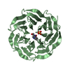
| #1: Protein | Mass: 34390.992 Da / Num. of mol.: 1 Source method: isolated from a genetically manipulated source Source: (gene. exp.) Homo sapiens (human) / Gene: WDR5, BIG3 / Production host:  | ||||
|---|---|---|---|---|---|
| #2: Chemical | ChemComp-ARG /   Type: L-peptide linking / Mass: 175.209 Da / Num. of mol.: 1 / Source method: obtained synthetically / Formula: C6H15N4O2 Type: L-peptide linking / Mass: 175.209 Da / Num. of mol.: 1 / Source method: obtained synthetically / Formula: C6H15N4O2 | ||||
| #3: Chemical |   Mass: 96.063 Da / Num. of mol.: 2 / Source method: obtained synthetically / Formula: SO4 Mass: 96.063 Da / Num. of mol.: 2 / Source method: obtained synthetically / Formula: SO4#4: Chemical | ChemComp-GOL /   Mass: 92.094 Da / Num. of mol.: 4 / Source method: obtained synthetically / Formula: C3H8O3 Mass: 92.094 Da / Num. of mol.: 4 / Source method: obtained synthetically / Formula: C3H8O3#5: Water | ChemComp-HOH / |  Mass: 18.015 Da / Num. of mol.: 243 / Source method: isolated from a natural source / Formula: H2O Mass: 18.015 Da / Num. of mol.: 243 / Source method: isolated from a natural source / Formula: H2O |
-Experimental details
-Experiment
| Experiment | Method: X-RAY DIFFRACTION / Number of used crystals: 1 |
|---|
- Sample preparation
| Crystal | Density Matthews: 2.1 Å3/Da / Density % sol: 40.4 % |
|---|---|
| Crystal grow | Temperature: 295 K / Method: vapor diffusion, sitting drop / pH: 5.9 Details: 100 mM Bis-Tris pH 5.9; 32% PEG3350, 54.6 mM Ammonium Sulfate, 0.66 mM L-arginine |
-Data collection
| Diffraction | Mean temperature: 100 K / Serial crystal experiment: N |
|---|---|
| Diffraction source | Source: SYNCHROTRON / Site: APS  / Beamline: 31-ID / Wavelength: 0.97931 Å / Beamline: 31-ID / Wavelength: 0.97931 Å |
| Detector | Type: RAYONIX MX225HE / Detector: CCD / Date: Oct 24, 2018 / Details: KB mirror |
| Radiation | Monochromator: diamond (111) / Protocol: SINGLE WAVELENGTH / Monochromatic (M) / Laue (L): M / Scattering type: x-ray |
| Radiation wavelength | Wavelength: 0.97931 Å / Relative weight: 1 |
| Reflection | Resolution: 1.92→59.26 Å / Num. obs: 22592 / % possible obs: 99 % / Redundancy: 12.5 % / Rpim(I) all: 0.04 / Net I/σ(I): 10.7 |
| Reflection shell | Resolution: 1.92→1.97 Å / Redundancy: 10.9 % / Mean I/σ(I) obs: 3.1 / Num. unique obs: 1272 / Rpim(I) all: 0.29 / % possible all: 84.7 |
- Processing
| Software |
| ||||||||||||||||||||||||||||||||||||||||||||||||||||||||||||||||||||||||||||||||||||||||||||||||||||||||||||||||||||||||||||||||||||||||||||||||||||||||||||||||||||||||||||||||||||||
|---|---|---|---|---|---|---|---|---|---|---|---|---|---|---|---|---|---|---|---|---|---|---|---|---|---|---|---|---|---|---|---|---|---|---|---|---|---|---|---|---|---|---|---|---|---|---|---|---|---|---|---|---|---|---|---|---|---|---|---|---|---|---|---|---|---|---|---|---|---|---|---|---|---|---|---|---|---|---|---|---|---|---|---|---|---|---|---|---|---|---|---|---|---|---|---|---|---|---|---|---|---|---|---|---|---|---|---|---|---|---|---|---|---|---|---|---|---|---|---|---|---|---|---|---|---|---|---|---|---|---|---|---|---|---|---|---|---|---|---|---|---|---|---|---|---|---|---|---|---|---|---|---|---|---|---|---|---|---|---|---|---|---|---|---|---|---|---|---|---|---|---|---|---|---|---|---|---|---|---|---|---|---|---|
| Refinement | Method to determine structure: MOLECULAR REPLACEMENT Starting model: 2H14 Resolution: 1.92→59.26 Å / Cor.coef. Fo:Fc: 0.97 / Cor.coef. Fo:Fc free: 0.955 / SU B: 3.493 / SU ML: 0.099 / Cross valid method: THROUGHOUT / ESU R: 0.154 / ESU R Free: 0.138 / Details: HYDROGENS HAVE BEEN ADDED IN THE RIDING POSITIONS
| ||||||||||||||||||||||||||||||||||||||||||||||||||||||||||||||||||||||||||||||||||||||||||||||||||||||||||||||||||||||||||||||||||||||||||||||||||||||||||||||||||||||||||||||||||||||
| Solvent computation | Ion probe radii: 0.8 Å / Shrinkage radii: 0.8 Å / VDW probe radii: 1.2 Å | ||||||||||||||||||||||||||||||||||||||||||||||||||||||||||||||||||||||||||||||||||||||||||||||||||||||||||||||||||||||||||||||||||||||||||||||||||||||||||||||||||||||||||||||||||||||
| Displacement parameters | Biso mean: 25.994 Å2
| ||||||||||||||||||||||||||||||||||||||||||||||||||||||||||||||||||||||||||||||||||||||||||||||||||||||||||||||||||||||||||||||||||||||||||||||||||||||||||||||||||||||||||||||||||||||
| Refinement step | Cycle: 1 / Resolution: 1.92→59.26 Å
| ||||||||||||||||||||||||||||||||||||||||||||||||||||||||||||||||||||||||||||||||||||||||||||||||||||||||||||||||||||||||||||||||||||||||||||||||||||||||||||||||||||||||||||||||||||||
| Refine LS restraints |
|