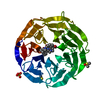
[English] 日本語
Yorodumi
- PDB-3emh: Structural basis of WDR5-MLL interaction -

+
Open data


ID or keywords:

Loading...

-
Basic information

Entry
Database: PDB / ID: 3emh
TitleStructural basis of WDR5-MLL interaction
Components
  • Mixed-lineage leukemia protein 1
  • WD repeat-containing protein 5
KeywordsGENE REGULATION / Chromatin / Histone / WD40 repeat / Nucleus / Phosphoprotein / WD repeat / Zinc-finger
Function / homology
Function and homology information


: / : / : / : / : / : / : / : / : / positive regulation of cellular response to drug ...: / : / : / : / : / : / : / : / : / positive regulation of cellular response to drug / [histone H3]-lysine4 N-trimethyltransferase / protein-cysteine methyltransferase activity / positive regulation of transporter activity / response to potassium ion / [histone H3]-lysine4 N-methyltransferase / histone H3K4 monomethyltransferase activity / regulation of megakaryocyte differentiation / unmethylated CpG binding / histone H3K4 trimethyltransferase activity / negative regulation of DNA methylation-dependent heterochromatin formation / T-helper 2 cell differentiation / MLL3/4 complex / regulation of short-term neuronal synaptic plasticity / Set1C/COMPASS complex / MLL1/2 complex / definitive hemopoiesis / ATAC complex / NSL complex / histone H3K4 methyltransferase activity / Cardiogenesis / regulation of hematopoietic stem cell differentiation / embryonic hemopoiesis / exploration behavior / anterior/posterior pattern specification / regulation of tubulin deacetylation / histone methyltransferase complex / Formation of WDR5-containing histone-modifying complexes / regulation of cell division / minor groove of adenine-thymine-rich DNA binding / membrane depolarization / regulation of embryonic development / MLL1 complex / histone acetyltransferase complex / negative regulation of fibroblast proliferation / homeostasis of number of cells within a tissue / positive regulation of gluconeogenesis / spleen development / lysine-acetylated histone binding / cellular response to transforming growth factor beta stimulus / methylated histone binding / transcription initiation-coupled chromatin remodeling / Transferases; Transferring one-carbon groups; Methyltransferases / post-embryonic development / skeletal system development / gluconeogenesis / Transcriptional regulation of granulopoiesis / RUNX1 regulates genes involved in megakaryocyte differentiation and platelet function / circadian regulation of gene expression / PKMTs methylate histone lysines / visual learning / protein modification process / RMTs methylate histone arginines / mitotic spindle / Activation of anterior HOX genes in hindbrain development during early embryogenesis / RUNX1 regulates transcription of genes involved in differentiation of HSCs / Neddylation / HATs acetylate histones / histone binding / fibroblast proliferation / protein-containing complex assembly / methylation / transcription cis-regulatory region binding / DNA-binding transcription factor activity, RNA polymerase II-specific / regulation of cell cycle / chromatin binding / regulation of transcription by RNA polymerase II / positive regulation of DNA-templated transcription / regulation of DNA-templated transcription / apoptotic process / negative regulation of transcription by RNA polymerase II / positive regulation of transcription by RNA polymerase II / protein homodimerization activity / zinc ion binding / nucleoplasm / identical protein binding / nucleus / cytosol
Similarity search - Function
Histone-lysine N-methyltransferase 2A / KMT2A, ePHD domain / KMT2A, PHD domain 1 / KMT2A, PHD domain 2 / KMT2A, PHD domain 3 / Methyltransferase, trithorax / : / FY-rich, N-terminal / F/Y-rich N-terminus / PHD-like zinc-binding domain ...Histone-lysine N-methyltransferase 2A / KMT2A, ePHD domain / KMT2A, PHD domain 1 / KMT2A, PHD domain 2 / KMT2A, PHD domain 3 / Methyltransferase, trithorax / : / FY-rich, N-terminal / F/Y-rich N-terminus / PHD-like zinc-binding domain / FYR domain FYRN motif profile. / "FY-rich" domain, N-terminal region / FY-rich, C-terminal / F/Y rich C-terminus / FYR domain FYRC motif profile. / "FY-rich" domain, C-terminal region / CXXC zinc finger domain / Zinc finger, CXXC-type / Zinc finger CXXC-type profile. / Cysteine-rich motif following a subset of SET domains / Post-SET domain / Post-SET domain profile. / SET (Su(var)3-9, Enhancer-of-zeste, Trithorax) domain / SET domain superfamily / SET domain / Extended PHD (ePHD) domain / Extended PHD (ePHD) domain profile. / SET domain profile. / SET domain / YVTN repeat-like/Quinoprotein amine dehydrogenase / 7 Propeller / Methylamine Dehydrogenase; Chain H / PHD-finger / Zinc finger PHD-type signature. / Zinc finger PHD-type profile. / Zinc finger, PHD-finger / Zinc finger, PHD-type / PHD zinc finger / Zinc finger, FYVE/PHD-type / Bromodomain profile. / bromo domain / Bromodomain / Bromodomain-like superfamily / G-protein beta WD-40 repeat / Zinc finger, RING/FYVE/PHD-type / WD40 repeat, conserved site / Trp-Asp (WD) repeats signature. / Trp-Asp (WD) repeats profile. / Trp-Asp (WD) repeats circular profile. / WD domain, G-beta repeat / WD40 repeats / WD40 repeat / WD40-repeat-containing domain superfamily / WD40/YVTN repeat-like-containing domain superfamily / Mainly Beta
Similarity search - Domain/homology
WD repeat-containing protein 5 / Histone-lysine N-methyltransferase 2A / Histone-lysine N-methyltransferase 2A
Similarity search - Component
Biological speciesHomo sapiens (human)
MethodX-RAY DIFFRACTION / SYNCHROTRON / MOLECULAR REPLACEMENT / Resolution: 1.37 Å
AuthorsSong, J.J. / Kingston, R.E.
CitationJournal: J.Biol.Chem. / Year: 2008
Title: WDR5 Interacts with Mixed Lineage Leukemia (MLL) Protein via the Histone H3-binding Pocket.
Authors: Song, J.J. / Kingston, R.E.
History
DepositionSep 24, 2008Deposition site: RCSB / Processing site: RCSB
Revision 1.0Oct 7, 2008Provider: repository / Type: Initial release
Revision 1.1Jul 13, 2011Group: Version format compliance
Revision 1.2Sep 6, 2023Group: Data collection / Database references ...Data collection / Database references / Derived calculations / Refinement description
Category: chem_comp_atom / chem_comp_bond ...chem_comp_atom / chem_comp_bond / database_2 / pdbx_initial_refinement_model / struct_ref_seq_dif / struct_site
Item: _database_2.pdbx_DOI / _database_2.pdbx_database_accession ..._database_2.pdbx_DOI / _database_2.pdbx_database_accession / _struct_ref_seq_dif.details / _struct_site.pdbx_auth_asym_id / _struct_site.pdbx_auth_comp_id / _struct_site.pdbx_auth_seq_id

-
Structure visualization

Structure viewerMolecule:
MolmilJmol/JSmol

Downloads & links

-
Assembly

Deposited unit
A: WD repeat-containing protein 5
B: Mixed-lineage leukemia protein 1
hetero molecules


Theoretical massNumber of molelcules
Total (without water)36,6124
Polymers36,4202
Non-polymers1922
Water4,810267
1


  • Idetical with deposited unit
  • defined by software
TypeNameSymmetry operationNumber
identity operation1_555x,y,z1
Buried area1010 Å2
ΔGint-5.2 kcal/mol
Surface area11580 Å2
MethodPISA
Unit cell
Length a, b, c (Å)78.440, 98.290, 80.030
Angle α, β, γ (deg.)90.00, 90.00, 90.00
Int Tables number20
Space group name H-MC2221
Components on special symmetry positions
IDModelComponents
11A-580-

HOH

-
Components

#1: Protein WD repeat-containing protein 5 / BMP2-induced 3-kb gene protein


Mass: 34916.590 Da / Num. of mol.: 1 / Fragment: UNP residues 25-334
Source method: isolated from a genetically manipulated source
Source: (gene. exp.) Homo sapiens (human) / Gene: WDR5, BIG3 / Plasmid: pGEX6p_1 / Production host: Escherichia coli (E. coli) / Strain (production host): BL21(DE3)pLysS / References: UniProt: P61964
#2: Protein/peptide Mixed-lineage leukemia protein 1 / MLL1


Mass: 1503.705 Da / Num. of mol.: 1 / Fragment: UNP residues 2675-2687 / Source method: obtained synthetically
Details: Synthetic peptide following the sequence position 2675-2687 of UniProt entry Q59FF2 (Q59FF2_HUMAN)
References: UniProt: Q59FF2, UniProt: Q03164*PLUS
#3: Chemical ChemComp-SO4 / SULFATE ION


Mass: 96.063 Da / Num. of mol.: 2 / Source method: obtained synthetically / Formula: SO4
#4: Water ChemComp-HOH / water


Mass: 18.015 Da / Num. of mol.: 267 / Source method: isolated from a natural source / Formula: H2O

-
Experimental details

-
Experiment

ExperimentMethod: X-RAY DIFFRACTION / Number of used crystals: 1

-
Sample preparation

CrystalDensity Matthews: 2.12 Å3/Da / Density % sol: 41.96 %
Crystal growTemperature: 293 K / Method: vapor diffusion, hanging drop / pH: 7.5
Details: 20 mM HEPES, 15-20% PEG 3350, 60mM Ammonium sulfate, pH 7.5, VAPOR DIFFUSION, HANGING DROP, temperature 293K

-
Data collection

DiffractionMean temperature: 110 K
Diffraction sourceSource: SYNCHROTRON / Site: NSLS / Beamline: X29A / Wavelength: 1.0809 Å
DetectorType: ADSC QUANTUM 315 / Detector: CCD / Date: Apr 29, 2008
RadiationMonochromator: 100 um / Protocol: SINGLE WAVELENGTH / Monochromatic (M) / Laue (L): M / Scattering type: x-ray
Radiation wavelengthWavelength: 1.0809 Å / Relative weight: 1
ReflectionResolution: 1.37→50 Å / Num. all: 64701 / Num. obs: 65115 / % possible obs: 99 % / Redundancy: 7.4 % / Rmerge(I) obs: 0.059 / Net I/σ(I): 36.1
Reflection shellResolution: 1.37→1.41 Å / Rmerge(I) obs: 0.25 / Mean I/σ(I) obs: 3.1 / % possible all: 90.3

-
Processing

Software
NameVersionClassification
HKL-2000data collection
PHASERphasing
CNS1.1refinement
HKL-2000data reduction
HKL-2000data scaling
RefinementMethod to determine structure: MOLECULAR REPLACEMENT
Starting model: PDB entry 2H13
Resolution: 1.37→50 Å / σ(F): 0 / Stereochemistry target values: Engh & Huber
RfactorNum. reflection
Rfree0.1998 3198
Rwork0.1903 -
obs0.1903 64701
all-64701
Refinement stepCycle: LAST / Resolution: 1.37→50 Å
ProteinNucleic acidLigandSolventTotal
Num. atoms2399 0 10 267 2676
Refine LS restraints
Refine-IDTypeDev ideal
X-RAY DIFFRACTIONc_bond_d0.004589
X-RAY DIFFRACTIONc_angle_deg1.89777
LS refinement shellResolution: 1.37→1.42 Å /
Num. reflection% reflection
obs5828 99 %

+
About Yorodumi

-
News

-
Feb 9, 2022. New format data for meta-information of EMDB entries

New format data for meta-information of EMDB entries

  • Version 3 of the EMDB header file is now the official format.
  • The previous official version 1.9 will be removed from the archive.

Related info.:EMDB header

External links:wwPDB to switch to version 3 of the EMDB data model

-
Aug 12, 2020. Covid-19 info

Covid-19 info

URL: https://pdbjlvh1.pdbj.org/emnavi/covid19.php

New page: Covid-19 featured information page in EM Navigator.

Related info.:Covid-19 info / Mar 5, 2020. Novel coronavirus structure data

+
Mar 5, 2020. Novel coronavirus structure data

Novel coronavirus structure data

Related info.:Yorodumi Speices / Aug 12, 2020. Covid-19 info

External links:COVID-19 featured content - PDBj / Molecule of the Month (242):Coronavirus Proteases

+
Jan 31, 2019. EMDB accession codes are about to change! (news from PDBe EMDB page)

EMDB accession codes are about to change! (news from PDBe EMDB page)

  • The allocation of 4 digits for EMDB accession codes will soon come to an end. Whilst these codes will remain in use, new EMDB accession codes will include an additional digit and will expand incrementally as the available range of codes is exhausted. The current 4-digit format prefixed with “EMD-” (i.e. EMD-XXXX) will advance to a 5-digit format (i.e. EMD-XXXXX), and so on. It is currently estimated that the 4-digit codes will be depleted around Spring 2019, at which point the 5-digit format will come into force.
  • The EM Navigator/Yorodumi systems omit the EMD- prefix.

Related info.:Q: What is EMD? / ID/Accession-code notation in Yorodumi/EM Navigator

External links:EMDB Accession Codes are Changing Soon! / Contact to PDBj

+
Jul 12, 2017. Major update of PDB

Major update of PDB

  • wwPDB released updated PDB data conforming to the new PDBx/mmCIF dictionary.
  • This is a major update changing the version number from 4 to 5, and with Remediation, in which all the entries are updated.
  • In this update, many items about electron microscopy experimental information are reorganized (e.g. em_software).
  • Now, EM Navigator and Yorodumi are based on the updated data.

External links:wwPDB Remediation / Enriched Model Files Conforming to OneDep Data Standards Now Available in the PDB FTP Archive

-
Yorodumi

Thousand views of thousand structures

  • Yorodumi is a browser for structure data from EMDB, PDB, SASBDB, etc.
  • This page is also the successor to EM Navigator detail page, and also detail information page/front-end page for Omokage search.
  • The word "yorodu" (or yorozu) is an old Japanese word meaning "ten thousand". "mi" (miru) is to see.

Related info.:EMDB / PDB / SASBDB / Comparison of 3 databanks / Yorodumi Search / Aug 31, 2016. New EM Navigator & Yorodumi / Yorodumi Papers / Jmol/JSmol / Function and homology information / Changes in new EM Navigator and Yorodumi

Read more