 Movie
Movie Controller

[English] 日本語
 Yorodumi
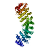
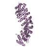
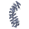
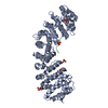
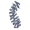
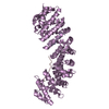
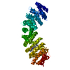
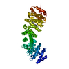
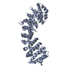
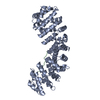
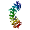
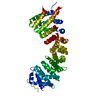
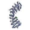
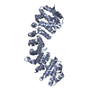
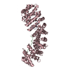
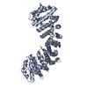
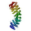
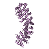
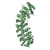
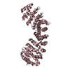
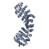
- PDB-3wpt: Crystal structure of closed dimer of human importin-alpha1 (Rch1) -
+ Open data

- Basic information
| Entry | Database: PDB / ID: 3wpt | ||||||
|---|---|---|---|---|---|---|---|
| Title | Crystal structure of closed dimer of human importin-alpha1 (Rch1) | ||||||
 Components Components | Importin subunit alpha-1 | ||||||
 Keywords Keywords | TRANSPORT PROTEIN / Arm repeat / All Alpha protein / Armadillo repeat / Closed dimer / Nuclear transport / importin-beta / NLS-carring proteins | ||||||
| Function / homology |  Function and homology information Function and homology informationSensing of DNA Double Strand Breaks / regulation of DNA recombination / entry of viral genome into host nucleus through nuclear pore complex via importin / positive regulation of viral life cycle / NS1 Mediated Effects on Host Pathways / NLS-dependent protein nuclear import complex / host cell / nuclear import signal receptor activity / CREB1 phosphorylation through the activation of CaMKII/CaMKK/CaMKIV cascasde / nuclear localization sequence binding ...Sensing of DNA Double Strand Breaks / regulation of DNA recombination / entry of viral genome into host nucleus through nuclear pore complex via importin / positive regulation of viral life cycle / NS1 Mediated Effects on Host Pathways / NLS-dependent protein nuclear import complex / host cell / nuclear import signal receptor activity / CREB1 phosphorylation through the activation of CaMKII/CaMKK/CaMKIV cascasde / nuclear localization sequence binding / DNA metabolic process / CaMK IV-mediated phosphorylation of CREB / NLS-bearing protein import into nucleus / ISG15 antiviral mechanism / protein import into nucleus / histone deacetylase binding / SARS-CoV-1 activates/modulates innate immune responses / nuclear membrane / Estrogen-dependent gene expression / Golgi membrane / endoplasmic reticulum membrane / SARS-CoV-2 activates/modulates innate and adaptive immune responses / RNA binding / nucleoplasm / membrane / nucleus / cytosol / cytoplasmSimilarity search - Function | ||||||
| Biological species |  Homo sapiens (human) Homo sapiens (human) | ||||||
| Method | X-RAY DIFFRACTION / SYNCHROTRON / MOLECULAR REPLACEMENT / Resolution: 2.629 Å | ||||||
 Authors Authors | Miyatake, H. / Sanjoh, A. / Matsuda, G. / Tatsumi, Y. / Dohmae, N. / Aida, Y. | ||||||
 Citation Citation | Journal: To be Published Title: Crystal structure of closed dimer of human importin-alpha1 (Rch1) Authors: Miyatake, H. | ||||||
| History |
|
- Structure visualization
| Structure viewer | Molecule: MolmilJmol/JSmol |
|---|
- Downloads & links
-Download
| PDBx/mmCIF format | 3wpt.cif.gz | 476.5 KB | Display | PDBx/mmCIF format |
|---|---|---|---|---|
| PDB format | pdb3wpt.ent.gz | 405.7 KB | Display | PDB format |
| PDBx/mmJSON format | 3wpt.json.gz | Tree view | PDBx/mmJSON format | |
| Others |  Other downloads Other downloads |
-Validation report
| Arichive directory | https://data.pdbj.org/pub/pdb/validation_reports/wp/3wptftp://data.pdbj.org/pub/pdb/validation_reports/wp/3wpt | HTTPS FTP |
|---|
-Related structure data
| Related structure data | 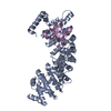 2jdqS  3wp7 S: Starting model for refinement |
|---|---|
| Similar structure data |

-Links
 PDBj
PDBj


- Assembly
| Deposited unit | 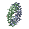
| ||||||||
|---|---|---|---|---|---|---|---|---|---|
| 1 | 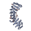
| ||||||||
| 2 | 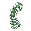
| ||||||||
| Unit cell |
|
-Components
| #1: Protein | / Rch1 / Karyopherin subunit alpha-2 / RAG cohort protein 1 / SRP1-alpha Mass: 45773.355 Da / Num. of mol.: 2 / Fragment: UNP residues 75-497 Source method: isolated from a genetically manipulated source Source: (gene. exp.) Homo sapiens (human) / Gene: KPNA2, RCH1, SRP1 / Production host:  Escherichia coli (E. coli) / References: UniProt: P52292 Escherichia coli (E. coli) / References: UniProt: P52292#2: Chemical | ChemComp-PEG / Diethylene glycol  Mass: 106.120 Da / Num. of mol.: 17 / Source method: obtained synthetically / Formula: C4H10O3 Mass: 106.120 Da / Num. of mol.: 17 / Source method: obtained synthetically / Formula: C4H10O3#3: Water | ChemComp-HOH / | Water Mass: 18.015 Da / Num. of mol.: 199 / Source method: isolated from a natural source / Formula: H2O Mass: 18.015 Da / Num. of mol.: 199 / Source method: isolated from a natural source / Formula: H2O |
|---|
-Experimental details
-Experiment
| Experiment | Method: X-RAY DIFFRACTION / Number of used crystals: 1 |
|---|
- Sample preparation
| Crystal | Density Matthews: 3.74 Å3/Da / Density % sol: 67.14 % |
|---|---|
| Crystal grow | Temperature: 293 K / Method: vapor diffusion, sitting drop / pH: 5.5 Details: 50MM MES, 100MM AMMONIUM SULFATE, 10MM MGCL, 15-20%(w/v) PEG8000, pH 5.5, VAPOR DIFFUSION, SITTING DROP, temperature 293K |
-Data collection
| Diffraction | Mean temperature: 100 K |
|---|---|
| Diffraction source | Source: SYNCHROTRON / Site: SPring-8  / Beamline: BL32B2 / Wavelength: 1 Å / Beamline: BL32B2 / Wavelength: 1 Å |
| Detector | Type: MARMOSAIC 225 mm CCD / Detector: CCD / Date: Jun 23, 2008 / Details: Mirror |
| Radiation | Monochromator: MIRRORS / Protocol: SINGLE WAVELENGTH / Monochromatic (M) / Laue (L): M / Scattering type: x-ray |
| Radiation wavelength | Wavelength: 1 Å / Relative weight: 1 |
| Reflection | Resolution: 2.629→46.51 Å / Num. obs: 40635 / % possible obs: 96.96 % / Observed criterion σ(F): 0 / Observed criterion σ(I): 0 / Redundancy: 8.7 % / Biso Wilson estimate: 48.38 Å2 / Rmerge(I) obs: 0.065 / Rsym value: 0.065 / Net I/σ(I): 31.83 |
| Reflection shell | Resolution: 2.629→2.723 Å / Redundancy: 4.8 % / Rmerge(I) obs: 0.391 / Mean I/σ(I) obs: 4.86 / Num. unique all: 3701 / Rsym value: 0.391 / % possible all: 90.25 |
- Processing
| Software |
| |||||||||||||||||||||||||||||||||||||||||||||||||||||||||||||||||||||||||||||||||||||||||||||||||||||||||||||||||||||||||||||||||||||||||||||||||||||||||||||||||||||||||||||||
|---|---|---|---|---|---|---|---|---|---|---|---|---|---|---|---|---|---|---|---|---|---|---|---|---|---|---|---|---|---|---|---|---|---|---|---|---|---|---|---|---|---|---|---|---|---|---|---|---|---|---|---|---|---|---|---|---|---|---|---|---|---|---|---|---|---|---|---|---|---|---|---|---|---|---|---|---|---|---|---|---|---|---|---|---|---|---|---|---|---|---|---|---|---|---|---|---|---|---|---|---|---|---|---|---|---|---|---|---|---|---|---|---|---|---|---|---|---|---|---|---|---|---|---|---|---|---|---|---|---|---|---|---|---|---|---|---|---|---|---|---|---|---|---|---|---|---|---|---|---|---|---|---|---|---|---|---|---|---|---|---|---|---|---|---|---|---|---|---|---|---|---|---|---|---|---|---|
| Refinement | Method to determine structure: MOLECULAR REPLACEMENT Starting model: 2JDQ Resolution: 2.629→46.505 Å / FOM work R set: 0.8636 / SU ML: 0.3 / σ(F): 1.49 / σ(I): 0 / Phase error: 20.79 / Stereochemistry target values: ML
| |||||||||||||||||||||||||||||||||||||||||||||||||||||||||||||||||||||||||||||||||||||||||||||||||||||||||||||||||||||||||||||||||||||||||||||||||||||||||||||||||||||||||||||||
| Solvent computation | Shrinkage radii: 0.9 Å / VDW probe radii: 1.11 Å / Solvent model: FLAT BULK SOLVENT MODEL | |||||||||||||||||||||||||||||||||||||||||||||||||||||||||||||||||||||||||||||||||||||||||||||||||||||||||||||||||||||||||||||||||||||||||||||||||||||||||||||||||||||||||||||||
| Displacement parameters | Biso max: 159.17 Å2 / Biso mean: 55.14 Å2 / Biso min: 23.91 Å2 | |||||||||||||||||||||||||||||||||||||||||||||||||||||||||||||||||||||||||||||||||||||||||||||||||||||||||||||||||||||||||||||||||||||||||||||||||||||||||||||||||||||||||||||||
| Refinement step | Cycle: LAST / Resolution: 2.629→46.505 Å
| |||||||||||||||||||||||||||||||||||||||||||||||||||||||||||||||||||||||||||||||||||||||||||||||||||||||||||||||||||||||||||||||||||||||||||||||||||||||||||||||||||||||||||||||
| Refine LS restraints |
| |||||||||||||||||||||||||||||||||||||||||||||||||||||||||||||||||||||||||||||||||||||||||||||||||||||||||||||||||||||||||||||||||||||||||||||||||||||||||||||||||||||||||||||||
| LS refinement shell | Refine-ID: X-RAY DIFFRACTION / Total num. of bins used: 14
| |||||||||||||||||||||||||||||||||||||||||||||||||||||||||||||||||||||||||||||||||||||||||||||||||||||||||||||||||||||||||||||||||||||||||||||||||||||||||||||||||||||||||||||||
| Refinement TLS params. | Method: refined / Refine-ID: X-RAY DIFFRACTION
| |||||||||||||||||||||||||||||||||||||||||||||||||||||||||||||||||||||||||||||||||||||||||||||||||||||||||||||||||||||||||||||||||||||||||||||||||||||||||||||||||||||||||||||||
| Refinement TLS group |
|
