 Movie
Movie Controller

[English] 日本語
 Yorodumi
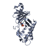
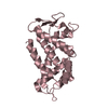
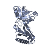
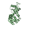
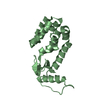
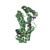
- PDB-3b76: Crystal structure of the third PDZ domain of human ligand-of-numb... -
+ Open data

- Basic information
| Entry | Database: PDB / ID: 3b76 | ||||||
|---|---|---|---|---|---|---|---|
| Title | Crystal structure of the third PDZ domain of human ligand-of-numb protein-X (LNX1) in complex with the C-terminal peptide from the coxsackievirus and adenovirus receptor | ||||||
 Components Components | E3 ubiquitin-protein ligase LNX | ||||||
 Keywords Keywords | LIGASE / PDZ / bound ligand / Structural Genomics / Structural Genomics Consortium / SGC / Metal-binding / Ubl conjugation pathway / Zinc-finger / UNKNOWN FUNCTION | ||||||
| Function / homology |  Function and homology information Function and homology informationPDZ domain binding / RING-type E3 ubiquitin transferase / ubiquitin-protein transferase activity / Antigen processing: Ubiquitination & Proteasome degradation / ubiquitin-dependent protein catabolic process / protein ubiquitination / identical protein binding / metal ion binding / cytoplasm Similarity search - Function | ||||||
| Biological species |  Homo sapiens (human) Homo sapiens (human) | ||||||
| Method |  X-RAY DIFFRACTION / SYNCHROTRON / MOLECULAR REPLACEMENT / Resolution: 1.75 Å X-RAY DIFFRACTION / SYNCHROTRON / MOLECULAR REPLACEMENT / Resolution: 1.75 Å | ||||||
 Authors Authors | Ugochukwu, E. / Burgess-Brown, N. / Berridge, G. / Elkins, J. / Bunkoczi, G. / Pike, A.C.W. / Sundstrom, M. / Arrowsmith, C.H. / Weigelt, J. / Edwards, A.M. ...Ugochukwu, E. / Burgess-Brown, N. / Berridge, G. / Elkins, J. / Bunkoczi, G. / Pike, A.C.W. / Sundstrom, M. / Arrowsmith, C.H. / Weigelt, J. / Edwards, A.M. / Gileadi, O. / von Delft, F. / Doyle, D. / Structural Genomics Consortium (SGC) | ||||||
 Citation Citation | Journal: To be Published Title: Crystal structure of the third PDZ domain of human ligand-of-numb protein-X (LNX1) in complex with the C-terminal peptide from the coxsackievirus and adenovirus receptor. Authors: Ugochukwu, E. / Burgess-Brown, N. / Berridge, G. / Elkins, J. / Bunkoczi, G. / Pike, A.C.W. / Sundstrom, M. / Arrowsmith, C.H. / Weigelt, J. / Edwards, A.M. / Gileadi, O. / von Delft, F. / Doyle, D. | ||||||
| History |
| ||||||
| Remark 999 | SEQUENCE This is a C-terminal peptide from the coxsackievirus and adenovirus receptor. |
- Structure visualization
| Structure viewer | Molecule: MolmilJmol/JSmol |
|---|
- Downloads & links
-Download
| PDBx/mmCIF format | 3b76.cif.gz | 57.6 KB | Display | PDBx/mmCIF format |
|---|---|---|---|---|
| PDB format | pdb3b76.ent.gz | 40 KB | Display | PDB format |
| PDBx/mmJSON format | 3b76.json.gz | Tree view | PDBx/mmJSON format | |
| Others |  Other downloads Other downloads |
-Validation report
| Summary document | 3b76_validation.pdf.gz | 451.4 KB | Display | wwPDB validaton report |
|---|---|---|---|---|
| Full document | 3b76_full_validation.pdf.gz | 451.6 KB | Display | |
| Data in XML | 3b76_validation.xml.gz | 10.9 KB | Display | |
| Data in CIF | 3b76_validation.cif.gz | 14.6 KB | Display | |
| Arichive directory | https://data.pdbj.org/pub/pdb/validation_reports/b7/3b76ftp://data.pdbj.org/pub/pdb/validation_reports/b7/3b76 | HTTPS FTP |
-Related structure data
| Related structure data |  2awuS 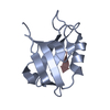 2awwS  2awxS  2bygS  2g2lS S: Starting model for refinement |
|---|---|
| Similar structure data |


-Links
 PDBj
PDBj

- Assembly
| Deposited unit | 
| ||||||||||||||||||
|---|---|---|---|---|---|---|---|---|---|---|---|---|---|---|---|---|---|---|---|
| 1 |
| ||||||||||||||||||
| Unit cell |
| ||||||||||||||||||
| Noncrystallographic symmetry (NCS) | NCS domain:
NCS domain segments: Component-ID: 1 / Ens-ID: 1 / Beg auth comp-ID: LEU / Beg label comp-ID: LEU / End auth comp-ID: VAL / End label comp-ID: VAL / Refine code: 5 / Auth seq-ID: 402 - 502 / Label seq-ID: 18 - 118
|
-Components
| #1: Protein | Mass: 12824.500 Da / Num. of mol.: 2 / Fragment: Third PDZ domain: Residues 504-594 Source method: isolated from a genetically manipulated source Source: (gene. exp.) Homo sapiens (human) / Gene: LNX1, LNX / Plasmid: pNIC28-Bsa4 / Production host:  References: UniProt: Q8TBB1, Ligases; Forming carbon-nitrogen bonds; Acid-amino-acid ligases (peptide synthases) #2: Chemical | ChemComp-NA / |   Mass: 22.990 Da / Num. of mol.: 1 / Source method: obtained synthetically / Formula: Na Mass: 22.990 Da / Num. of mol.: 1 / Source method: obtained synthetically / Formula: Na#3: Chemical |   Mass: 62.068 Da / Num. of mol.: 2 / Source method: obtained synthetically / Formula: C2H6O2 Mass: 62.068 Da / Num. of mol.: 2 / Source method: obtained synthetically / Formula: C2H6O2#4: Water | ChemComp-HOH / |  Mass: 18.015 Da / Num. of mol.: 124 / Source method: isolated from a natural source / Formula: H2O Mass: 18.015 Da / Num. of mol.: 124 / Source method: isolated from a natural source / Formula: H2O |
|---|
-Experimental details
-Experiment
| Experiment | Method: X-RAY DIFFRACTION / Number of used crystals: 1 |
|---|
- Sample preparation
| Crystal | Density Matthews: 2.09 Å3/Da / Density % sol: 41.19 % |
|---|---|
| Crystal grow | Temperature: 293 K / Method: vapor diffusion, sitting drop / pH: 7.4 Details: 1.53M Potassium phosphate, 0.27M Sodium phosphate, 1% PEG 3350, pH 7.4, VAPOR DIFFUSION, SITTING DROP, temperature 293K |
-Data collection
| Diffraction | Mean temperature: 100 K |
|---|---|
| Diffraction source | Source: SYNCHROTRON / Site: SLS  / Beamline: X10SA / Wavelength: 0.99975 Å / Beamline: X10SA / Wavelength: 0.99975 Å |
| Detector | Type: MARMOSAIC 225 mm CCD / Detector: CCD / Date: Jun 18, 2007 |
| Radiation | Protocol: SINGLE WAVELENGTH / Monochromatic (M) / Laue (L): M / Scattering type: x-ray |
| Radiation wavelength | Wavelength: 0.99975 Å / Relative weight: 1 |
| Reflection | Resolution: 1.75→33.71 Å / Num. all: 22359 / Num. obs: 22359 / % possible obs: 100 % / Observed criterion σ(F): 0 / Observed criterion σ(I): 0 / Redundancy: 7.2 % / Biso Wilson estimate: 20.4 Å2 / Rmerge(I) obs: 0.094 / Rsym value: 0.094 / Net I/σ(I): 16 |
| Reflection shell | Resolution: 1.75→1.84 Å / Redundancy: 7.3 % / Rmerge(I) obs: 0.844 / Mean I/σ(I) obs: 2.9 / Num. unique all: 23287 / Rsym value: 0.844 / % possible all: 100 |
- Processing
| Software |
| |||||||||||||||||||||||||||||||||||||||||||||||||||||||||||||||||||||||||||||||||||||||||||||||||||||||||||||||||||||||||||||
|---|---|---|---|---|---|---|---|---|---|---|---|---|---|---|---|---|---|---|---|---|---|---|---|---|---|---|---|---|---|---|---|---|---|---|---|---|---|---|---|---|---|---|---|---|---|---|---|---|---|---|---|---|---|---|---|---|---|---|---|---|---|---|---|---|---|---|---|---|---|---|---|---|---|---|---|---|---|---|---|---|---|---|---|---|---|---|---|---|---|---|---|---|---|---|---|---|---|---|---|---|---|---|---|---|---|---|---|---|---|---|---|---|---|---|---|---|---|---|---|---|---|---|---|---|---|---|
| Refinement | Method to determine structure: MOLECULAR REPLACEMENT Starting model: PDB entries 2AWW, 2BYG, 2AWX, 2AWU, 2G2L Resolution: 1.75→33.71 Å / Cor.coef. Fo:Fc: 0.96 / Cor.coef. Fo:Fc free: 0.944 / SU B: 4.124 / SU ML: 0.067 / TLS residual ADP flag: LIKELY RESIDUAL / Cross valid method: THROUGHOUT / σ(F): 0 / ESU R: 0.105 / ESU R Free: 0.102 / Stereochemistry target values: MAXIMUM LIKELIHOOD / Details: HYDROGENS HAVE BEEN ADDED IN THE RIDING POSITIONS
| |||||||||||||||||||||||||||||||||||||||||||||||||||||||||||||||||||||||||||||||||||||||||||||||||||||||||||||||||||||||||||||
| Solvent computation | Ion probe radii: 0.8 Å / Shrinkage radii: 0.8 Å / VDW probe radii: 1.2 Å / Solvent model: MASK | |||||||||||||||||||||||||||||||||||||||||||||||||||||||||||||||||||||||||||||||||||||||||||||||||||||||||||||||||||||||||||||
| Displacement parameters | Biso mean: 17.652 Å2
| |||||||||||||||||||||||||||||||||||||||||||||||||||||||||||||||||||||||||||||||||||||||||||||||||||||||||||||||||||||||||||||
| Refinement step | Cycle: LAST / Resolution: 1.75→33.71 Å
| |||||||||||||||||||||||||||||||||||||||||||||||||||||||||||||||||||||||||||||||||||||||||||||||||||||||||||||||||||||||||||||
| Refine LS restraints |
| |||||||||||||||||||||||||||||||||||||||||||||||||||||||||||||||||||||||||||||||||||||||||||||||||||||||||||||||||||||||||||||
| Refine LS restraints NCS | Ens-ID: 1 / Refine-ID: X-RAY DIFFRACTION
| |||||||||||||||||||||||||||||||||||||||||||||||||||||||||||||||||||||||||||||||||||||||||||||||||||||||||||||||||||||||||||||
| LS refinement shell | Resolution: 1.75→1.795 Å / Total num. of bins used: 20
| |||||||||||||||||||||||||||||||||||||||||||||||||||||||||||||||||||||||||||||||||||||||||||||||||||||||||||||||||||||||||||||
| Refinement TLS params. | Method: refined / Refine-ID: X-RAY DIFFRACTION
| |||||||||||||||||||||||||||||||||||||||||||||||||||||||||||||||||||||||||||||||||||||||||||||||||||||||||||||||||||||||||||||
| Refinement TLS group |
|
