 Movie
Movie Controller

[English] 日本語
 Yorodumi
- PDB-4ann: Crystal Structure Staphylococcus aureus ESSB cytoplasmic fragment -
+ Open data

- Basic information
| Entry | Database: PDB / ID: 4ann | ||||||
|---|---|---|---|---|---|---|---|
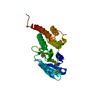
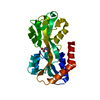
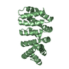
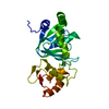
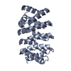
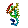
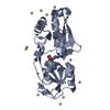
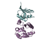
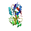
| Title | Crystal Structure Staphylococcus aureus ESSB cytoplasmic fragment | ||||||
 Components Components | ESSB | ||||||
 Keywords Keywords | MEMBRANE PROTEIN / MEMBRANE SECRETION / ESS TYPE VII SECRETION SYSTEM / VIRULENCE | ||||||
| Function / homology | Type VII secretion system EssB / Type VII secretion system EssB, C-terminal / WXG100 protein secretion system (Wss), protein YukC / Transferase(Phosphotransferase) domain 1 / Transferase(Phosphotransferase); domain 1 / Orthogonal Bundle / Mainly Alpha / plasma membrane / Type VII secretion system protein EssB Function and homology information Function and homology information | ||||||
| Biological species |  STAPHYLOCOCCUS AUREUS SUBSP. AUREUS NCTC 8325 (bacteria) STAPHYLOCOCCUS AUREUS SUBSP. AUREUS NCTC 8325 (bacteria) | ||||||
| Method |  X-RAY DIFFRACTION / SYNCHROTRON / SAD / Resolution: 1.05 Å X-RAY DIFFRACTION / SYNCHROTRON / SAD / Resolution: 1.05 Å | ||||||
 Authors Authors | Zoltner, M. / Fyfe, P.K. / Palmer, T. / Hunter, W.N. | ||||||
 Citation Citation | Journal: Biochem.J. / Year: 2013 Title: Characterization of Staphylococcus Aureus Essb, an Integral Membrane Component of the Type Vii Secretion System: Atomic Resolution Crystal Structure of the Cytoplasmic Segment. Authors: Zoltner, M. / Fyfe, P.K. / Palmer, T. / Hunter, W.N. | ||||||
| History |
| ||||||
| Remark 650 | HELIX DETERMINATION METHOD: AUTHOR PROVIDED. | ||||||
| Remark 700 | SHEET DETERMINATION METHOD: AUTHOR PROVIDED. |
- Structure visualization
| Structure viewer | Molecule: MolmilJmol/JSmol |
|---|
- Downloads & links
-Download
| PDBx/mmCIF format | 4ann.cif.gz | 105.4 KB | Display | PDBx/mmCIF format |
|---|---|---|---|---|
| PDB format | pdb4ann.ent.gz | 81.4 KB | Display | PDB format |
| PDBx/mmJSON format | 4ann.json.gz | Tree view | PDBx/mmJSON format | |
| Others |  Other downloads Other downloads |
-Validation report
| Arichive directory | https://data.pdbj.org/pub/pdb/validation_reports/an/4annftp://data.pdbj.org/pub/pdb/validation_reports/an/4ann | HTTPS FTP |
|---|
-Related structure data
| Related structure data | |
|---|---|
| Similar structure data |


-Links
 PDBj
PDBj- Assembly
| Deposited unit | 
| ||||||||
|---|---|---|---|---|---|---|---|---|---|
| 1 |
| ||||||||
| Unit cell |
|
-Components
| #1: Protein | Mass: 25123.406 Da / Num. of mol.: 1 / Fragment: CYTOPLASMIC FRAGMENT, RESIDUES 12-226 Source method: isolated from a genetically manipulated source Source: (gene. exp.) STAPHYLOCOCCUS AUREUS SUBSP. AUREUS NCTC 8325 (bacteria)Plasmid: MODIFIED PET27 / Production host: |
|---|---|
| #2: Water | ChemComp-HOH /  Mass: 18.015 Da / Num. of mol.: 199 / Source method: isolated from a natural source / Formula: H2O Mass: 18.015 Da / Num. of mol.: 199 / Source method: isolated from a natural source / Formula: H2O |
-Experimental details
-Experiment
| Experiment | Method: X-RAY DIFFRACTION / Number of used crystals: 1 |
|---|
- Sample preparation
| Crystal | Density Matthews: 2.28 Å3/Da / Density % sol: 46.1 % / Description: NONE |
|---|---|
| Crystal grow | pH: 5 Details: 0.18 M AMMONIUM CITRATE PH5.0, 20 % (W/V) PEG3350, 0.25 MM TRIS(2-CARBOXYETHYL)PHOSPHINEHYDROCHLORIDE, 5% GLYCEROL |
-Data collection
| Diffraction | Mean temperature: 293 K |
|---|---|
| Diffraction source | Source: SYNCHROTRON / Site: ESRF  / Beamline: ID29 / Wavelength: 0.9814 / Beamline: ID29 / Wavelength: 0.9814 |
| Detector | Type: ADSC QUANTUM 315r / Detector: CCD / Date: May 14, 2009 / Details: MIRRORS |
| Radiation | Monochromator: SILICON / Protocol: SINGLE WAVELENGTH / Monochromatic (M) / Laue (L): M / Scattering type: x-ray |
| Radiation wavelength | Wavelength: 0.9814 Å / Relative weight: 1 |
| Reflection | Resolution: 1.05→58.74 Å / Num. obs: 100245 / % possible obs: 99 % / Observed criterion σ(I): 0 / Redundancy: 7.1 % / Rmerge(I) obs: 0.06 / Net I/σ(I): 18 |
| Reflection shell | Resolution: 1.05→1.15 Å / Redundancy: 4.6 % / Rmerge(I) obs: 0.41 / Mean I/σ(I) obs: 3.6 / % possible all: 92.8 |
- Processing
| Software |
| |||||||||||||||||||||||||||||||||
|---|---|---|---|---|---|---|---|---|---|---|---|---|---|---|---|---|---|---|---|---|---|---|---|---|---|---|---|---|---|---|---|---|---|---|
| Refinement | Method to determine structure: SAD Starting model: NONE Resolution: 1.05→50 Å / Num. parameters: 17570 / Num. restraintsaints: 25556 / Cross valid method: FREE R-VALUE / σ(F): 0 / Stereochemistry target values: ENGH AND HUBER Details: ANISOTROPIC REFINEMENT REDUCED FREE R (NO CUTOFF) BY 3.0 PERCENT (FINAL RFREE 0.158)
| |||||||||||||||||||||||||||||||||
| Solvent computation | Solvent model: MOEWS & KRETSINGER | |||||||||||||||||||||||||||||||||
| Refine analyze | Num. disordered residues: 201 / Occupancy sum hydrogen: 1420.97 / Occupancy sum non hydrogen: 1636.02 | |||||||||||||||||||||||||||||||||
| Refinement step | Cycle: LAST / Resolution: 1.05→50 Å
| |||||||||||||||||||||||||||||||||
| Refine LS restraints |
|
