 Movie
Movie Controller

[English] 日本語
 Yorodumi
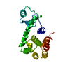
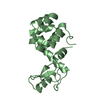
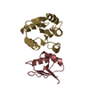
- PDB-2g3v: Crystal structure of CagS (HP0534, Cag13) from Helicobacter pylori -
+ Open data

- Basic information
| Entry | Database: PDB / ID: 2g3v | ||||||
|---|---|---|---|---|---|---|---|
| Title | Crystal structure of CagS (HP0534, Cag13) from Helicobacter pylori | ||||||
 Components Components |
| ||||||
 Keywords Keywords | UNKNOWN FUNCTION / Helicobacter pylori / pathogenicity island / Type IV secretion system | ||||||
| Function / homology | CAG pathogenicity island protein 13, CagS / CAG pathogenicity island protein 13 / CagS superfamily / Cag pathogenicity island protein S of Helicobacter pylori / Four Helix Bundle (Hemerythrin (Met), subunit A) / Up-down Bundle / Mainly Alpha / CAG pathogenicity island protein 13 Function and homology information Function and homology information | ||||||
| Biological species |   Helicobacter pylori (bacteria) Helicobacter pylori (bacteria) | ||||||
| Method |  X-RAY DIFFRACTION / SYNCHROTRON / MAD / Resolution: 2.3 Å X-RAY DIFFRACTION / SYNCHROTRON / MAD / Resolution: 2.3 Å | ||||||
 Authors Authors | Cendron, L. / Tasca, E. / Angelini, A. / Seydel, A. / Battistutta, R. / Montecucco, C. / Zanotti, G. | ||||||
 Citation Citation | Journal: To be Published Title: Crystal structure of CagS from helicobacter pylori Authors: Cendron, L. / Tasca, E. / Angelini, A. / Seydel, A. / Battistutta, R. / Montecucco, C. / Zanotti, G. #1: Journal: J.Mol.Biol. / Year: 2004Title: Crystal structure of CagZ, a protein from the Helicobacter pylori pathogenicity island that encodes for a type IV secretion system Authors: Cendron, L. / Seydel, A. / Angelini, A. / Battistutta, R. / Zanotti, G. | ||||||
| History |
|
- Structure visualization
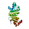
| Structure viewer | Molecule: MolmilJmol/JSmol |
|---|
- Downloads & links
-Download
| PDBx/mmCIF format | 2g3v.cif.gz | 151.4 KB | Display | PDBx/mmCIF format |
|---|---|---|---|---|
| PDB format | pdb2g3v.ent.gz | 122.7 KB | Display | PDB format |
| PDBx/mmJSON format | 2g3v.json.gz | Tree view | PDBx/mmJSON format | |
| Others |  Other downloads Other downloads |
-Validation report
| Arichive directory | https://data.pdbj.org/pub/pdb/validation_reports/g3/2g3vftp://data.pdbj.org/pub/pdb/validation_reports/g3/2g3v | HTTPS FTP |
|---|
-Related structure data
| Similar structure data |
|---|


-Links
 PDBj
PDBj
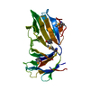
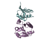
- Assembly
| Deposited unit | 
| ||||||||
|---|---|---|---|---|---|---|---|---|---|
| 1 | 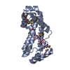
| ||||||||
| 2 | 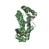
| ||||||||
| 3 | 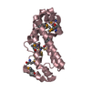
| ||||||||
| 4 | 
| ||||||||
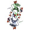
| Unit cell |
|
-Components
| #1: Protein | Mass: 25536.822 Da / Num. of mol.: 4 Source method: isolated from a genetically manipulated source Source: (gene. exp.) Helicobacter pylori (bacteria) / Strain: G27 / Gene: cagS, cag13, HP0534 / Plasmid: pET21b / Species (production host): Escherichia coli / Production host: #2: Protein/peptide | ( Mass: 706.735 Da / Num. of mol.: 4 / Source method: obtained synthetically / Details: The peptide was chemically synthesized. #3: Water | ChemComp-HOH / |  Mass: 18.015 Da / Num. of mol.: 238 / Source method: isolated from a natural source / Formula: H2O Mass: 18.015 Da / Num. of mol.: 238 / Source method: isolated from a natural source / Formula: H2OHas protein modification | Y | |
|---|
-Experimental details
-Experiment
| Experiment | Method: X-RAY DIFFRACTION / Number of used crystals: 3 |
|---|
- Sample preparation
| Crystal | Density Matthews: 2.2 Å3/Da / Density % sol: 43.2 % |
|---|---|
| Crystal grow | Temperature: 283 K / Method: vapor diffusion, hanging drop / pH: 8.5 Details: 0.2M KBr, 25% PEG 2000 MME, 0.1M Tris pH 8.5 , VAPOR DIFFUSION, HANGING DROP, temperature 283K |
-Data collection
| Diffraction | Mean temperature: 100 K | |||||||||||||||
|---|---|---|---|---|---|---|---|---|---|---|---|---|---|---|---|---|
| Diffraction source | Source: SYNCHROTRON / Site: ESRF  / Beamline: ID29 / Wavelength: 0.9792, 0.9794, 0.9756, 1.0 / Beamline: ID29 / Wavelength: 0.9792, 0.9794, 0.9756, 1.0 | |||||||||||||||
| Detector | Type: ADSC QUANTUM 210 / Detector: CCD / Date: Mar 12, 2005 / Details: Mirrors | |||||||||||||||
| Radiation | Monochromator: Si(311) / Protocol: MAD / Monochromatic (M) / Laue (L): M / Scattering type: x-ray | |||||||||||||||
| Radiation wavelength |
| |||||||||||||||
| Reflection | Resolution: 2.3→51 Å / Num. all: 38297 / Num. obs: 38297 / % possible obs: 99.9 % / Observed criterion σ(F): 0 / Observed criterion σ(I): 0 / Redundancy: 4.8 % / Biso Wilson estimate: 6.7 Å2 / Rsym value: 0.011 / Net I/σ(I): 5 | |||||||||||||||
| Reflection shell | Resolution: 2.3→2.42 Å / Redundancy: 4.9 % / Mean I/σ(I) obs: 1.7 / Num. unique all: 5569 / Rsym value: 0.41 / % possible all: 100 |
- Processing
| Software |
| ||||||||||||||||||||||||||||||||||||
|---|---|---|---|---|---|---|---|---|---|---|---|---|---|---|---|---|---|---|---|---|---|---|---|---|---|---|---|---|---|---|---|---|---|---|---|---|---|
| Refinement | Method to determine structure: MAD / Resolution: 2.3→39.41 Å / Rfactor Rfree error: 0.006 / Data cutoff high absF: 2250186.93 / Data cutoff low absF: 0 / Isotropic thermal model: RESTRAINED / Cross valid method: THROUGHOUT / σ(F): 0 / Stereochemistry target values: Engh & Huber
| ||||||||||||||||||||||||||||||||||||
| Solvent computation | Solvent model: FLAT MODEL / Bsol: 56.1075 Å2 / ksol: 0.40001 e/Å3 | ||||||||||||||||||||||||||||||||||||
| Displacement parameters | Biso mean: 10.4 Å2
| ||||||||||||||||||||||||||||||||||||
| Refine analyze |
| ||||||||||||||||||||||||||||||||||||
| Refinement step | Cycle: LAST / Resolution: 2.3→39.41 Å
| ||||||||||||||||||||||||||||||||||||
| Refine LS restraints |
| ||||||||||||||||||||||||||||||||||||
| LS refinement shell | Resolution: 2.3→2.4 Å / Rfactor Rfree error: 0.015 / Total num. of bins used: 6
| ||||||||||||||||||||||||||||||||||||
| Xplor file |
|
