 Movie
Movie Controller


+ Open data

- Basic information
| Entry | Database: PDB / ID: 4ryn | ||||||
|---|---|---|---|---|---|---|---|
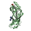
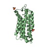
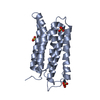
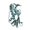
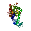
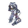
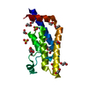
| Title | Crystal structure of BcTSPO, type1 monomer | ||||||
 Components Components | Integral membrane protein | ||||||
 Keywords Keywords | MEMBRANE PROTEIN / Structural Genomics / PSI-Biology / Protein Structure Initiative / New York Consortium on Membrane Protein Structure / NYCOMPS / Receptor | ||||||
| Function / homology |  Function and homology information Function and homology informationtetrapyrrole metabolic process / tetrapyrrole binding / membrane / identical protein binding / plasma membrane Similarity search - Function | ||||||
| Biological species |  | ||||||
| Method |  X-RAY DIFFRACTION / SYNCHROTRON / MOLECULAR REPLACEMENT / Resolution: 2.01 Å X-RAY DIFFRACTION / SYNCHROTRON / MOLECULAR REPLACEMENT / Resolution: 2.01 Å | ||||||
 Authors Authors | Guo, Y. / Liu, Q. / Hendrickson, W.A. / New York Consortium on Membrane Protein Structure (NYCOMPS) | ||||||
 Citation Citation | Journal: Science / Year: 2015 Title: Protein structure. Structure and activity of tryptophan-rich TSPO proteins. Authors: Guo, Y. / Kalathur, R.C. / Liu, Q. / Kloss, B. / Bruni, R. / Ginter, C. / Kloppmann, E. / Rost, B. / Hendrickson, W.A. | ||||||
| History |
|
- Structure visualization
| Structure viewer | Molecule: MolmilJmol/JSmol |
|---|
- Downloads & links
-Download
| PDBx/mmCIF format | 4ryn.cif.gz | 53.7 KB | Display | PDBx/mmCIF format |
|---|---|---|---|---|
| PDB format | pdb4ryn.ent.gz | 35.8 KB | Display | PDB format |
| PDBx/mmJSON format | 4ryn.json.gz | Tree view | PDBx/mmJSON format | |
| Others |  Other downloads Other downloads |
-Validation report
| Arichive directory | https://data.pdbj.org/pub/pdb/validation_reports/ry/4rynftp://data.pdbj.org/pub/pdb/validation_reports/ry/4ryn | HTTPS FTP |
|---|
-Related structure data
| Related structure data |  4ryiC  4ryjC  4rymSC  4ryoC  4ryqC  4ryrC C: citing same article ( S: Starting model for refinement |
|---|---|
| Similar structure data | |
| Other databases |


-Links
 PDBj
PDBj


- Assembly
| Deposited unit | 
| ||||||||
|---|---|---|---|---|---|---|---|---|---|
| 1 |
| ||||||||
| Unit cell |
|
-Components
-Protein / Sugars , 2 types, 2 molecules A
| #1: Protein | Mass: 21496.965 Da / Num. of mol.: 1 Source method: isolated from a genetically manipulated source Source: (gene. exp.) |
|---|---|
| #3: Sugar | ChemComp-LMU /  Type: D-saccharide / Mass: 510.615 Da / Num. of mol.: 1 / Source method: obtained synthetically / Formula: C24H46O11 / Comment: detergent*YM Type: D-saccharide / Mass: 510.615 Da / Num. of mol.: 1 / Source method: obtained synthetically / Formula: C24H46O11 / Comment: detergent*YM |
-Non-polymers , 4 types, 38 molecules 


| #2: Chemical | ChemComp-MPG / [(  Mass: 356.540 Da / Num. of mol.: 9 / Source method: obtained synthetically / Formula: C21H40O4 Mass: 356.540 Da / Num. of mol.: 9 / Source method: obtained synthetically / Formula: C21H40O4#4: Chemical | ChemComp-CAC / |  Mass: 136.989 Da / Num. of mol.: 1 / Source method: obtained synthetically / Formula: C2H6AsO2 Mass: 136.989 Da / Num. of mol.: 1 / Source method: obtained synthetically / Formula: C2H6AsO2#5: Chemical | ChemComp-PGE / |  Mass: 150.173 Da / Num. of mol.: 1 / Source method: obtained synthetically / Formula: C6H14O4 Mass: 150.173 Da / Num. of mol.: 1 / Source method: obtained synthetically / Formula: C6H14O4#6: Water | ChemComp-HOH / | Mass: 18.015 Da / Num. of mol.: 27 / Source method: isolated from a natural source / Formula: H2O |
|---|
-Experimental details
-Experiment
| Experiment | Method: X-RAY DIFFRACTION / Number of used crystals: 1 |
|---|
- Sample preparation
| Crystal | Density Matthews: 1.94 Å3/Da / Density % sol: 36.66 % |
|---|---|
| Crystal grow | Temperature: 293 K / Method: lcp / pH: 6.5 Details: crystals grew from 0.1 M sodium cacodylate, 5% w/v PGA LM (poly-l-glutamic acid, low molecular weight~ 200-400 kDa), 30% v/v PEG 550MME (Polyethylene glycol monomethyl ether 550), pH 6.5 in LCP, temperature 293K |
-Data collection
| Diffraction | Mean temperature: 100 K |
|---|---|
| Diffraction source | Source: SYNCHROTRON / Site: NSLS  / Beamline: X4C / Wavelength: 0.9791 Å / Beamline: X4C / Wavelength: 0.9791 Å |
| Detector | Type: MAR CCD 165 mm / Detector: CCD / Date: Apr 3, 2014 |
| Radiation | Monochromator: Single crystal bender / Protocol: SINGLE WAVELENGTH / Monochromatic (M) / Laue (L): M / Scattering type: x-ray |
| Radiation wavelength | Wavelength: 0.9791 Å / Relative weight: 1 |
| Reflection | Resolution: 2.01→49 Å / Num. obs: 11360 / % possible obs: 99.4 % / Observed criterion σ(F): 0 / Observed criterion σ(I): 0 / Redundancy: 11.6 % / Rmerge(I) obs: 0.23062 / Net I/σ(I): 3.3 |
| Reflection shell | Resolution: 2.01→2.06 Å / % possible all: 95.8 |
- Processing
| Software |
| |||||||||||||||||||||||||||||||||||
|---|---|---|---|---|---|---|---|---|---|---|---|---|---|---|---|---|---|---|---|---|---|---|---|---|---|---|---|---|---|---|---|---|---|---|---|---|
| Refinement | Method to determine structure: MOLECULAR REPLACEMENT Starting model: PDB ENTRY 4RYM Resolution: 2.01→34.754 Å / SU ML: 0.25 / σ(F): 1.34 / Phase error: 29.58 / Stereochemistry target values: ML
| |||||||||||||||||||||||||||||||||||
| Solvent computation | Shrinkage radii: 0.9 Å / VDW probe radii: 1.11 Å / Solvent model: FLAT BULK SOLVENT MODEL | |||||||||||||||||||||||||||||||||||
| Refinement step | Cycle: LAST / Resolution: 2.01→34.754 Å
| |||||||||||||||||||||||||||||||||||
| Refine LS restraints |
| |||||||||||||||||||||||||||||||||||
| LS refinement shell |
|
