 Movie
Movie Controller


+ Open data

- Basic information
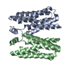
| Entry | Database: PDB / ID: 4ryi | ||||||
|---|---|---|---|---|---|---|---|
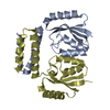
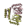
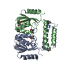
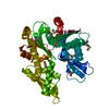
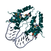
| Title | Crystal structure of BcTSPO/PK11195 complex | ||||||
 Components Components | Integral membrane protein | ||||||
 Keywords Keywords | MEMBRANE PROTEIN / Structural Genomics / PSI-Biology / Protein Structure Initiative / New York Consortium on Membrane Protein Structure / NYCOMPS / Receptor | ||||||
| Function / homology |  Function and homology information Function and homology informationtetrapyrrole metabolic process / tetrapyrrole binding / membrane / identical protein binding / plasma membrane Similarity search - Function | ||||||
| Biological species |  | ||||||
| Method |  X-RAY DIFFRACTION / SYNCHROTRON / MOLECULAR REPLACEMENT / Resolution: 3.49 Å X-RAY DIFFRACTION / SYNCHROTRON / MOLECULAR REPLACEMENT / Resolution: 3.49 Å | ||||||
 Authors Authors | Guo, Y. / Liu, Q. / Hendrickson, W.A. / New York Consortium on Membrane Protein Structure (NYCOMPS) | ||||||
 Citation Citation | Journal: Science / Year: 2015 Title: Protein structure. Structure and activity of tryptophan-rich TSPO proteins. Authors: Guo, Y. / Kalathur, R.C. / Liu, Q. / Kloss, B. / Bruni, R. / Ginter, C. / Kloppmann, E. / Rost, B. / Hendrickson, W.A. | ||||||
| History |
|
- Structure visualization
| Structure viewer | Molecule: MolmilJmol/JSmol |
|---|
- Downloads & links
-Download
| PDBx/mmCIF format | 4ryi.cif.gz | 74.5 KB | Display | PDBx/mmCIF format |
|---|---|---|---|---|
| PDB format | pdb4ryi.ent.gz | 56.5 KB | Display | PDB format |
| PDBx/mmJSON format | 4ryi.json.gz | Tree view | PDBx/mmJSON format | |
| Others |  Other downloads Other downloads |
-Validation report
| Arichive directory | https://data.pdbj.org/pub/pdb/validation_reports/ry/4ryiftp://data.pdbj.org/pub/pdb/validation_reports/ry/4ryi | HTTPS FTP |
|---|
-Related structure data
| Related structure data |  4ryjC  4rymC  4rynC  4ryoC  4ryqC  4ryrC C: citing same article ( |
|---|---|
| Similar structure data | |
| Other databases |
-Links
 PDBj
PDBj- Assembly
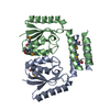
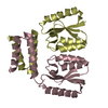
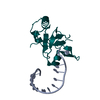
| Deposited unit | 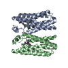
| ||||||||
|---|---|---|---|---|---|---|---|---|---|
| 1 |
| ||||||||
| 2 | 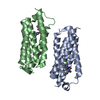
| ||||||||
| Unit cell |
|
-Components
| #1: Protein | Mass: 21496.965 Da / Num. of mol.: 2 Source method: isolated from a genetically manipulated source Source: (gene. exp.) #2: Chemical | 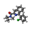  Mass: 352.857 Da / Num. of mol.: 2 / Source method: obtained synthetically / Formula: C21H21ClN2O Mass: 352.857 Da / Num. of mol.: 2 / Source method: obtained synthetically / Formula: C21H21ClN2O |
|---|
-Experimental details
-Experiment
| Experiment | Method: X-RAY DIFFRACTION / Number of used crystals: 1 |
|---|
- Sample preparation
| Crystal | Density Matthews: 2.03 Å3/Da / Density % sol: 39.31 % |
|---|---|
| Crystal grow | Temperature: 293 K / pH: 7.5 Details: 3% PEG 4000, 0.066 sodium chloride, 0.02 Tris,pH 7.5 as crystallization buffer, covered the drop of a mix of (10mg/mL protein and saturated PK11195 in 8% DMSO):monoolein in a ratio of 2/3 ...Details: 3% PEG 4000, 0.066 sodium chloride, 0.02 Tris,pH 7.5 as crystallization buffer, covered the drop of a mix of (10mg/mL protein and saturated PK11195 in 8% DMSO):monoolein in a ratio of 2/3 (v/v), LCP, temperature 293K |
-Data collection
| Diffraction | Mean temperature: 100 K |
|---|---|
| Diffraction source | Source: SYNCHROTRON / Site: NSLS  / Beamline: X4C / Wavelength: 0.9791 Å / Beamline: X4C / Wavelength: 0.9791 Å |
| Detector | Type: MAR CCD 165 mm / Detector: CCD / Date: Jun 21, 2014 |
| Radiation | Monochromator: Single crystal bender / Protocol: SINGLE WAVELENGTH / Monochromatic (M) / Laue (L): M / Scattering type: x-ray |
| Radiation wavelength | Wavelength: 0.9791 Å / Relative weight: 1 |
| Reflection | Resolution: 3.49→50 Å / Num. obs: 4245 / % possible obs: 96.2 % / Observed criterion σ(F): 0 / Observed criterion σ(I): 0 / Redundancy: 3.2 % / Rmerge(I) obs: 0.176 / Net I/σ(I): 4.35 |
| Reflection shell | Resolution: 3.5→3.56 Å / % possible all: 88.6 |
- Processing
| Software |
| |||||||||||||||||||||||||
|---|---|---|---|---|---|---|---|---|---|---|---|---|---|---|---|---|---|---|---|---|---|---|---|---|---|---|
| Refinement | Method to determine structure: MOLECULAR REPLACEMENT / Resolution: 3.49→35.12 Å / SU ML: 0.4 / σ(F): 1.34 / Phase error: 36.5 / Stereochemistry target values: ML
| |||||||||||||||||||||||||
| Solvent computation | Shrinkage radii: 0.9 Å / VDW probe radii: 1.11 Å / Solvent model: FLAT BULK SOLVENT MODEL | |||||||||||||||||||||||||
| Refinement step | Cycle: LAST / Resolution: 3.49→35.12 Å
| |||||||||||||||||||||||||
| Refine LS restraints |
| |||||||||||||||||||||||||
| LS refinement shell | Resolution: 3.49→35.1214 Å
|
