 Movie
Movie Controller

[English] 日本語
 Yorodumi
- PDB-2xch: Crystal structure of PDK1 in complex with a pyrazoloquinazoline i... -
+ Open data

- Basic information
| Entry | Database: PDB / ID: 2xch | ||||||
|---|---|---|---|---|---|---|---|
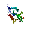
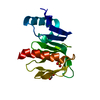
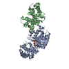
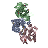
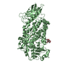
| Title | Crystal structure of PDK1 in complex with a pyrazoloquinazoline inhibitor | ||||||
 Components Components | 3-PHOSPHOINOSITIDE DEPENDENT PROTEIN KINASE 1 | ||||||
 Keywords Keywords | TRANSFERASE / PI3-KINASE SIGNALLING / SERINE/THREONINE-PROTEIN KINASE / ATP-BINDING | ||||||
| Function / homology |  Function and homology information Function and homology informationintracellular signaling cassette / 3-phosphoinositide-dependent protein kinase activity / Activation of AKT2 / regulation of mast cell degranulation / type B pancreatic cell development / negative regulation of toll-like receptor signaling pathway / RSK activation / positive regulation of vascular endothelial cell proliferation / regulation of canonical NF-kappaB signal transduction / hyperosmotic response ...intracellular signaling cassette / 3-phosphoinositide-dependent protein kinase activity / Activation of AKT2 / regulation of mast cell degranulation / type B pancreatic cell development / negative regulation of toll-like receptor signaling pathway / RSK activation / positive regulation of vascular endothelial cell proliferation / regulation of canonical NF-kappaB signal transduction / hyperosmotic response / negative regulation of cardiac muscle cell apoptotic process / phospholipase activator activity / positive regulation of sprouting angiogenesis / Constitutive Signaling by AKT1 E17K in Cancer / positive regulation of blood vessel endothelial cell migration / CD28 dependent PI3K/Akt signaling / Role of LAT2/NTAL/LAB on calcium mobilization / negative regulation of endothelial cell apoptotic process / SARS-CoV-2 targets host intracellular signalling and regulatory pathways / Estrogen-stimulated signaling through PRKCZ / vascular endothelial cell response to laminar fluid shear stress / SARS-CoV-1 targets host intracellular signalling and regulatory pathways / phospholipase binding / GPVI-mediated activation cascade / RHO GTPases activate PKNs / T cell costimulation / extrinsic apoptotic signaling pathway / Integrin signaling / insulin-like growth factor receptor signaling pathway / cellular response to epidermal growth factor stimulus / positive regulation of release of sequestered calcium ion into cytosol / cell projection / VEGFR2 mediated cell proliferation / VEGFR2 mediated vascular permeability / positive regulation of protein localization to plasma membrane / phosphatidylinositol 3-kinase/protein kinase B signal transduction / negative regulation of transforming growth factor beta receptor signaling pathway / calcium-mediated signaling / CLEC7A (Dectin-1) signaling / epidermal growth factor receptor signaling pathway / FCERI mediated NF-kB activation / cellular response to insulin stimulus / positive regulation of angiogenesis / Regulation of TP53 Degradation / insulin receptor signaling pathway / G beta:gamma signalling through PI3Kgamma / cell migration / Downstream TCR signaling / PIP3 activates AKT signaling / protein autophosphorylation / actin cytoskeleton organization / cytoplasmic vesicle / High laminar flow shear stress activates signaling by PIEZO1 and PECAM1:CDH5:KDR in endothelial cells / protein phosphorylation / non-specific serine/threonine protein kinase / positive regulation of phosphatidylinositol 3-kinase/protein kinase B signal transduction / intracellular signal transduction / postsynaptic density / protein serine kinase activity / focal adhesion / protein serine/threonine kinase activity / ATP binding / nucleus / plasma membrane / cytoplasm / cytosol Similarity search - Function | ||||||
| Biological species |  HOMO SAPIENS (human) HOMO SAPIENS (human) | ||||||
| Method |  X-RAY DIFFRACTION / SYNCHROTRON / MOLECULAR REPLACEMENT / Resolution: 2 Å X-RAY DIFFRACTION / SYNCHROTRON / MOLECULAR REPLACEMENT / Resolution: 2 Å | ||||||
 Authors Authors | Angiolini, M. / Banfi, P. / Casale, E. / Casuscelli, F. / Fiorelli, C. / Saccardo, M.B. / Silvagni, M. / Zuccotto, F. | ||||||
 Citation Citation | Journal: Bioorg.Med.Chem.Lett. / Year: 2010 Title: Structure-Based Optimization of Potent Pdk1 Inhibitors. Authors: Angiolini, M. / Banfi, P. / Casale, E. / Casuscelli, F. / Fiorelli, C. / Saccardo, M.B. / Silvagni, M. / Zuccotto, F. | ||||||
| History |
|
- Structure visualization
| Structure viewer | Molecule: MolmilJmol/JSmol |
|---|
- Downloads & links
-Download
| PDBx/mmCIF format | 2xch.cif.gz | 77.8 KB | Display | PDBx/mmCIF format |
|---|---|---|---|---|
| PDB format | pdb2xch.ent.gz | 57.7 KB | Display | PDB format |
| PDBx/mmJSON format | 2xch.json.gz | Tree view | PDBx/mmJSON format | |
| Others |  Other downloads Other downloads |
-Validation report
| Arichive directory | https://data.pdbj.org/pub/pdb/validation_reports/xc/2xchftp://data.pdbj.org/pub/pdb/validation_reports/xc/2xch | HTTPS FTP |
|---|
-Related structure data
| Related structure data |  2xckC  1h1wS C: citing same article ( S: Starting model for refinement |
|---|---|
| Similar structure data |


-Links
 PDBj
PDBj


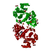
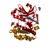
- Assembly
| Deposited unit | 
| ||||||||
|---|---|---|---|---|---|---|---|---|---|
| 1 | 
| ||||||||
| Unit cell |
|
-Components
| #1: Protein | Mass: 35427.609 Da / Num. of mol.: 1 / Fragment: KINASE CATALYTIC DOMAIN, RESIDUES 1-309 Source method: isolated from a genetically manipulated source Source: (gene. exp.) HOMO SAPIENS (human) / Production host:   SPODOPTERA FRUGIPERDA (fall armyworm) SPODOPTERA FRUGIPERDA (fall armyworm)References: UniProt: O15530, non-specific serine/threonine protein kinase | ||||||
|---|---|---|---|---|---|---|---|
| #2: Chemical | ChemComp-CKG /   Mass: 403.480 Da / Num. of mol.: 1 / Source method: obtained synthetically / Formula: C22H25N7O Mass: 403.480 Da / Num. of mol.: 1 / Source method: obtained synthetically / Formula: C22H25N7O | ||||||
| #3: Chemical | ChemComp-GOL /   Mass: 92.094 Da / Num. of mol.: 6 / Source method: obtained synthetically / Formula: C3H8O3 Mass: 92.094 Da / Num. of mol.: 6 / Source method: obtained synthetically / Formula: C3H8O3#4: Chemical | ChemComp-SO4 /   Mass: 96.063 Da / Num. of mol.: 6 / Source method: obtained synthetically / Formula: SO4 Mass: 96.063 Da / Num. of mol.: 6 / Source method: obtained synthetically / Formula: SO4#5: Water | ChemComp-HOH / |  Mass: 18.015 Da / Num. of mol.: 150 / Source method: isolated from a natural source / Formula: H2O Mass: 18.015 Da / Num. of mol.: 150 / Source method: isolated from a natural source / Formula: H2OHas protein modification | Y | |
-Experimental details
-Experiment
| Experiment | Method: X-RAY DIFFRACTION / Number of used crystals: 1 |
|---|
- Sample preparation
| Crystal | Density Matthews: 2.9 Å3/Da / Density % sol: 57.3 % / Description: NONE |
|---|---|
| Crystal grow | Details: 2.0 M AMMONIUM SULFATE,0.1 M TRIS PH8.5 |
-Data collection
| Diffraction | Mean temperature: 100 K |
|---|---|
| Diffraction source | Source: SYNCHROTRON / Site: ESRF  / Beamline: ID23-1 / Wavelength: 0.979 / Beamline: ID23-1 / Wavelength: 0.979 |
| Detector | Type: ADSC CCD / Detector: CCD / Date: Nov 27, 2006 |
| Radiation | Protocol: SINGLE WAVELENGTH / Monochromatic (M) / Laue (L): M / Scattering type: x-ray |
| Radiation wavelength | Wavelength: 0.979 Å / Relative weight: 1 |
| Reflection | Resolution: 2→30 Å / Num. obs: 27886 / % possible obs: 99.7 % / Observed criterion σ(I): 0 / Redundancy: 4.5 % / Rmerge(I) obs: 0.08 / Net I/σ(I): 9.8 |
| Reflection shell | Resolution: 2→2.1 Å / Redundancy: 4 % / Rmerge(I) obs: 0.39 / % possible all: 99.4 |
- Processing
| Software |
| ||||||||||||||||||||||||||||||||||||||||||||||||||||||||||||||||||||||||||||||||||||||||||||||||||||||||||||||||||||||||||||||||||||||||||||||||||||||||||||||||||||||||||||||||||||||
|---|---|---|---|---|---|---|---|---|---|---|---|---|---|---|---|---|---|---|---|---|---|---|---|---|---|---|---|---|---|---|---|---|---|---|---|---|---|---|---|---|---|---|---|---|---|---|---|---|---|---|---|---|---|---|---|---|---|---|---|---|---|---|---|---|---|---|---|---|---|---|---|---|---|---|---|---|---|---|---|---|---|---|---|---|---|---|---|---|---|---|---|---|---|---|---|---|---|---|---|---|---|---|---|---|---|---|---|---|---|---|---|---|---|---|---|---|---|---|---|---|---|---|---|---|---|---|---|---|---|---|---|---|---|---|---|---|---|---|---|---|---|---|---|---|---|---|---|---|---|---|---|---|---|---|---|---|---|---|---|---|---|---|---|---|---|---|---|---|---|---|---|---|---|---|---|---|---|---|---|---|---|---|---|
| Refinement | Method to determine structure: MOLECULAR REPLACEMENT Starting model: PDB ENTRY 1H1W Resolution: 2→30 Å / Cor.coef. Fo:Fc: 0.95 / Cor.coef. Fo:Fc free: 0.933 / SU B: 3.104 / SU ML: 0.09 / Cross valid method: THROUGHOUT / ESU R: 0.151 / ESU R Free: 0.142 / Stereochemistry target values: MAXIMUM LIKELIHOOD / Details: HYDROGENS HAVE BEEN ADDED IN THE RIDING POSITIONS.
| ||||||||||||||||||||||||||||||||||||||||||||||||||||||||||||||||||||||||||||||||||||||||||||||||||||||||||||||||||||||||||||||||||||||||||||||||||||||||||||||||||||||||||||||||||||||
| Solvent computation | Ion probe radii: 0.8 Å / Shrinkage radii: 0.8 Å / VDW probe radii: 1.4 Å / Solvent model: MASK | ||||||||||||||||||||||||||||||||||||||||||||||||||||||||||||||||||||||||||||||||||||||||||||||||||||||||||||||||||||||||||||||||||||||||||||||||||||||||||||||||||||||||||||||||||||||
| Displacement parameters | Biso mean: 26.771 Å2
| ||||||||||||||||||||||||||||||||||||||||||||||||||||||||||||||||||||||||||||||||||||||||||||||||||||||||||||||||||||||||||||||||||||||||||||||||||||||||||||||||||||||||||||||||||||||
| Refinement step | Cycle: LAST / Resolution: 2→30 Å
| ||||||||||||||||||||||||||||||||||||||||||||||||||||||||||||||||||||||||||||||||||||||||||||||||||||||||||||||||||||||||||||||||||||||||||||||||||||||||||||||||||||||||||||||||||||||
| Refine LS restraints |
|