Resolution: 1.6→50 Å / Num. obs: 107774 / % possible obs: 99.7 % / Redundancy: 13.2 % / Rmerge(I) obs: 0.107 / Χ2: 1.003 / Net I/σ(I): 8.4
Reflection shell
Resolution (Å)
Redundancy (%)
Rmerge(I) obs
Num. unique all
Χ2
Diffraction-ID
% possible all
1.6-1.63
11.6
0.995
5124
1
1
96.1
1.63-1.66
11.8
0.952
5256
0.999
1
98.2
1.66-1.69
12.1
0.732
5298
1.011
1
99.5
1.69-1.72
12.5
0.682
5335
1.007
1
99.8
1.72-1.76
12.9
0.664
5401
1.007
1
100
1.76-1.8
13
0.587
5323
1.009
1
100
1.8-1.85
13.1
0.444
5365
1.008
1
100
1.85-1.9
13.1
0.391
5394
1.029
1
100
1.9-1.95
13.2
0.281
5381
0.995
1
100
1.95-2.02
13.4
0.234
5352
0.97
1
100
2.02-2.09
13.5
0.197
5392
0.959
1
100
2.09-2.17
13.6
0.17
5378
0.965
1
100
2.17-2.27
13.6
0.16
5395
1.059
1
100
2.27-2.39
13.7
0.158
5404
1.135
1
100
2.39-2.54
13.7
0.156
5398
1.2
1
100
2.54-2.74
13.8
0.147
5427
1.171
1
100
2.74-3.01
13.8
0.121
5452
1.022
1
100
3.01-3.45
13.9
0.076
5449
0.774
1
100
3.45-4.34
13.8
0.059
5535
0.877
1
100
4.34-50
13.3
0.06
5715
0.876
1
99.7
-
Processing
Software
Name
Version
Classification
NB
SCALEPACK
datascaling
REFMAC
refinement
PDB_EXTRACT
3.11
dataextraction
HKL-2000
datareduction
SHELXS
phasing
Refinement
Method to determine structure: SAD / Resolution: 1.6→31.26 Å / Cor.coef. Fo:Fc: 0.966 / Cor.coef. Fo:Fc free: 0.959 / WRfactor Rfree: 0.2306 / WRfactor Rwork: 0.2129 / Occupancy max: 1 / Occupancy min: 0.3 / FOM work R set: 0.8014 / SU B: 3.749 / SU ML: 0.062 / SU R Cruickshank DPI: 0.0193 / SU Rfree: 0.0184 / Cross valid method: THROUGHOUT / σ(F): 0 / ESU R: 0.019 / ESU R Free: 0.018 / Stereochemistry target values: MAXIMUM LIKELIHOOD Details: U VALUES : WITH TLS ADDED HYDROGENS HAVE BEEN USED IF PRESENT IN THE INPUT
Rfactor
Num. reflection
% reflection
Selection details
Rfree
0.221
5372
5 %
RANDOM
Rwork
0.203
-
-
-
obs
0.204
107668
99.6 %
-
Solvent computation
Ion probe radii: 0.8 Å / Shrinkage radii: 0.8 Å / VDW probe radii: 1.2 Å / Solvent model: MASK
In the structure databanks used in Yorodumi, some data are registered as the other names, "COVID-19 virus" and "2019-nCoV". Here are the details of the virus and the list of structure data.
Jan 31, 2019. EMDB accession codes are about to change! (news from PDBe EMDB page)
EMDB accession codes are about to change! (news from PDBe EMDB page)
The allocation of 4 digits for EMDB accession codes will soon come to an end. Whilst these codes will remain in use, new EMDB accession codes will include an additional digit and will expand incrementally as the available range of codes is exhausted. The current 4-digit format prefixed with “EMD-” (i.e. EMD-XXXX) will advance to a 5-digit format (i.e. EMD-XXXXX), and so on. It is currently estimated that the 4-digit codes will be depleted around Spring 2019, at which point the 5-digit format will come into force.
The EM Navigator/Yorodumi systems omit the EMD- prefix.
Related info.:Q: What is EMD? / ID/Accession-code notation in Yorodumi/EM Navigator
Yorodumi is a browser for structure data from EMDB, PDB, SASBDB, etc.
This page is also the successor to EM Navigator detail page, and also detail information page/front-end page for Omokage search.
The word "yorodu" (or yorozu) is an old Japanese word meaning "ten thousand". "mi" (miru) is to see.
Related info.:EMDB / PDB / SASBDB / Comparison of 3 databanks / Yorodumi Search / Aug 31, 2016. New EM Navigator & Yorodumi / Yorodumi Papers / Jmol/JSmol / Function and homology information / Changes in new EM Navigator and Yorodumi
 Movie
Movie Controller

 Yorodumi
 Open data
 Basic information
 Components
 Keywords
 Function and homology information
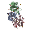
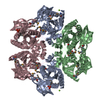
 Meiothermus ruber DSM 1279 (bacteria)
 X-RAY DIFFRACTION /
  Authors
 Citation
 Structure visualization
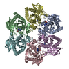
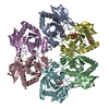
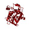
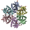
 Downloads & links
 Other downloads
 PDBj
PDBj Assembly
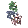

 Mass: 94.971 Da / Num. of mol.: 2 / Source method: obtained synthetically / Formula: PO4
 Mass: 24.305 Da / Num. of mol.: 1 / Source method: obtained synthetically / Formula: Mg
 Mass: 18.015 Da / Num. of mol.: 453 / Source method: isolated from a natural source / Formula: H2O
 Sample preparation
 / Beamline: 31-ID / Wavelength: 0.9791 Å
 Processing