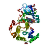
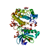
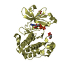
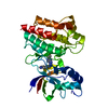
- PDB-1z5m: Crystal Structure Of N1-[3-[[5-bromo-2-[[3-[(1-pyrrolidinylcarbon... -
+
Open data
ID or keywords:
Loading...
-
Basic information
Entry
Database: PDB / ID: 1z5m
Title
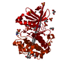
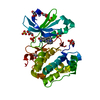
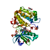
Crystal Structure Of N1-[3-[[5-bromo-2-[[3-[(1-pyrrolidinylcarbonyl)amino]phenyl]amino]-4-pyrimidinyl]amino]propyl]-2,2-dimethylpropanediamide Complexed with Human PDK1
Components
3-phosphoinositide dependent protein kinase-1
Keywords
TRANSFERASE / PROTEIN INHIBITOR COMPLEX / SERINE KINASE
Function / homology
Function and homology information
intracellular signaling cassette / 3-phosphoinositide-dependent protein kinase activity / Activation of AKT2 / regulation of mast cell degranulation / type B pancreatic cell development / negative regulation of toll-like receptor signaling pathway / RSK activation / regulation of canonical NF-kappaB signal transduction / hyperosmotic response / positive regulation of vascular endothelial cell proliferation ...intracellular signaling cassette / 3-phosphoinositide-dependent protein kinase activity / Activation of AKT2 / regulation of mast cell degranulation / type B pancreatic cell development / negative regulation of toll-like receptor signaling pathway / RSK activation / regulation of canonical NF-kappaB signal transduction / hyperosmotic response / positive regulation of vascular endothelial cell proliferation / phospholipase activator activity / positive regulation of sprouting angiogenesis / Constitutive Signaling by AKT1 E17K in Cancer / negative regulation of cardiac muscle cell apoptotic process / Role of LAT2/NTAL/LAB on calcium mobilization / CD28 dependent PI3K/Akt signaling / positive regulation of blood vessel endothelial cell migration / negative regulation of endothelial cell apoptotic process / SARS-CoV-2 targets host intracellular signalling and regulatory pathways / vascular endothelial cell response to laminar fluid shear stress / Estrogen-stimulated signaling through PRKCZ / SARS-CoV-1 targets host intracellular signalling and regulatory pathways / RHO GTPases activate PKNs / GPVI-mediated activation cascade / extrinsic apoptotic signaling pathway / phospholipase binding / T cell costimulation / Integrin signaling / cell projection / insulin-like growth factor receptor signaling pathway / cellular response to epidermal growth factor stimulus / ciliary tip / positive regulation of release of sequestered calcium ion into cytosol / VEGFR2 mediated cell proliferation / VEGFR2 mediated vascular permeability / positive regulation of protein localization to plasma membrane / phosphatidylinositol 3-kinase/protein kinase B signal transduction / negative regulation of transforming growth factor beta receptor signaling pathway / calcium-mediated signaling / CLEC7A (Dectin-1) signaling / epidermal growth factor receptor signaling pathway / FCERI mediated NF-kB activation / cellular response to insulin stimulus / positive regulation of angiogenesis / insulin receptor signaling pathway / Regulation of TP53 Degradation / protein autophosphorylation / PIP3 activates AKT signaling / Downstream TCR signaling / G beta:gamma signalling through PI3Kgamma / cell migration / actin cytoskeleton organization / cytoplasmic vesicle / High laminar flow shear stress activates signaling by PIEZO1 and PECAM1:CDH5:KDR in endothelial cells / protein phosphorylation / non-specific serine/threonine protein kinase / positive regulation of phosphatidylinositol 3-kinase/protein kinase B signal transduction / postsynaptic density / intracellular signal transduction / protein serine kinase activity / focal adhesion / protein serine/threonine kinase activity / ATP binding / nucleus / plasma membrane / cytoplasm / cytosol Similarity search - Function
PDK1-type, PH domain / PH domain / PDPK1 family / : / PH-like domain superfamily / Phosphorylase Kinase; domain 1 / Phosphorylase Kinase; domain 1 / Transferase(Phosphotransferase) domain 1 / Transferase(Phosphotransferase); domain 1 / Serine/threonine-protein kinase, active site ...PDK1-type, PH domain / PH domain / PDPK1 family / : / PH-like domain superfamily / Phosphorylase Kinase; domain 1 / Phosphorylase Kinase; domain 1 / Transferase(Phosphotransferase) domain 1 / Transferase(Phosphotransferase); domain 1 / Serine/threonine-protein kinase, active site / Serine/Threonine protein kinases active-site signature. / Protein kinase domain / Serine/Threonine protein kinases, catalytic domain / Protein kinase, ATP binding site / Protein kinases ATP-binding region signature. / Protein kinase domain profile. / Protein kinase domain / Protein kinase-like domain superfamily / 2-Layer Sandwich / Orthogonal Bundle / Mainly Alpha / Alpha Beta Similarity search - Domain/homology
HETEROGEN THE INHIBITION CONSTANT (KI APPARENT) FOR N-(3-{[5-BROMO-2-({3-[(PYRROLIDIN-1- YLCARBONYL) ...HETEROGEN THE INHIBITION CONSTANT (KI APPARENT) FOR N-(3-{[5-BROMO-2-({3-[(PYRROLIDIN-1- YLCARBONYL)AMINO]PHENYL}AMINO)PYRIMIDIN -4-YL]AMINO}PROPYL)-2,2-DIMETHYLMALONAMIDE AGAINST PDK1 WAS MEASURED AS 30 NM (N=5) DIRECT MEASURE PDK1 ACTIVITY WAS PERFORMED BY THE FOLLOWING PROTOCOL: THE FINAL ASSAY MIXTURE (60 UL) CONTAINED 50 MM TRIS- HCL, PH 7.5, 0.1 MM EGTA, 0.1 MM EDTA, 0.1% BETA-MERCAPTOETHANOL, 1 MG/ML BOVINE SERUM ALBUMIN, 10 MM MGOAC, 10 UM ATP, 0.2 UCI OF [GAMMA-33P]ATP, 7.5 UM OF SUBSTRATE PEPTIDE (H2NARRRGVTTKTFCGT) AND 60 NG OF PURIFIED RECOMBINANT HUMAN PDK1. AFTER 4 H AT ROOM TEMPERATURE, WE ADDED 25 MM EDTA AND SPOTTED A PORTION OF THE REACTION MIXTURE ON WHATMAN P81 PHOSPHOCELLULOSE PAPER. THE FILTER PAPER WAS WASHED 3 TIMES WITH 0.75% PHOSPHORIC ACID AND ONCE WITH ACETONE. AFTER DRYING, THE FILTER-BOUND LABELED PEPTIDE WAS QUANTIFIED USING A FUJI PHOSPHOIMAGER.
In the structure databanks used in Yorodumi, some data are registered as the other names, "COVID-19 virus" and "2019-nCoV". Here are the details of the virus and the list of structure data.
Jan 31, 2019. EMDB accession codes are about to change! (news from PDBe EMDB page)
EMDB accession codes are about to change! (news from PDBe EMDB page)
The allocation of 4 digits for EMDB accession codes will soon come to an end. Whilst these codes will remain in use, new EMDB accession codes will include an additional digit and will expand incrementally as the available range of codes is exhausted. The current 4-digit format prefixed with “EMD-” (i.e. EMD-XXXX) will advance to a 5-digit format (i.e. EMD-XXXXX), and so on. It is currently estimated that the 4-digit codes will be depleted around Spring 2019, at which point the 5-digit format will come into force.
The EM Navigator/Yorodumi systems omit the EMD- prefix.
Related info.:Q: What is EMD? / ID/Accession-code notation in Yorodumi/EM Navigator
Yorodumi is a browser for structure data from EMDB, PDB, SASBDB, etc.
This page is also the successor to EM Navigator detail page, and also detail information page/front-end page for Omokage search.
The word "yorodu" (or yorozu) is an old Japanese word meaning "ten thousand". "mi" (miru) is to see.
Related info.:EMDB / PDB / SASBDB / Comparison of 3 databanks / Yorodumi Search / Aug 31, 2016. New EM Navigator & Yorodumi / Yorodumi Papers / Jmol/JSmol / Function and homology information / Changes in new EM Navigator and Yorodumi
 Movie
Movie Controller

 Yorodumi
 Open data
 Basic information
 Components
 Keywords
 Function and homology information
 Homo sapiens (human)
 X-RAY DIFFRACTION /
  Authors
 Citation
 Structure visualization
 Downloads & links
 Other downloads
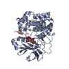


 PDBj
PDBj


 Assembly
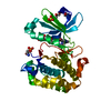
 unidentified baculovirus / Strain (production host): SF-21 cells / References: UniProt: O15530, EC: 2.7.1.37


 Mass: 96.063 Da / Num. of mol.: 1 / Source method: obtained synthetically / Formula: SO4
 Mass: 35.453 Da / Num. of mol.: 1 / Source method: obtained synthetically / Formula: Cl
 Mass: 547.448 Da / Num. of mol.: 1 / Source method: obtained synthetically / Formula: C23H31BrN8O3
 Mass: 92.094 Da / Num. of mol.: 3 / Source method: obtained synthetically / Formula: C3H8O3
 Sample preparation
 / Beamline: 5.0.1 / Wavelength: 1 Å
 Processing