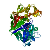
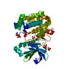
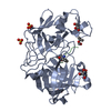
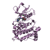
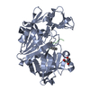
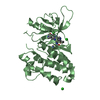
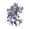
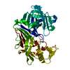
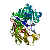
[English] 日本語
Yorodumi
- PDB-3nuu: phosphoinositide-dependent kinase-1 (PDK1) with fragment11 -

+
Open data


ID or keywords:

Loading...

-
Basic information

Entry
Database: PDB / ID: 3nuu
Titlephosphoinositide-dependent kinase-1 (PDK1) with fragment11
ComponentsPkB-like
KeywordsTRANSFERASE / Kinase domain
Function / homology
Function and homology information


3-phosphoinositide-dependent protein kinase activity / Activation of AKT2 / regulation of mast cell degranulation / negative regulation of toll-like receptor signaling pathway / type B pancreatic cell development / positive regulation of phospholipase activity / hyperosmotic response / RSK activation / regulation of canonical NF-kappaB signal transduction / positive regulation of vascular endothelial cell proliferation ...3-phosphoinositide-dependent protein kinase activity / Activation of AKT2 / regulation of mast cell degranulation / negative regulation of toll-like receptor signaling pathway / type B pancreatic cell development / positive regulation of phospholipase activity / hyperosmotic response / RSK activation / regulation of canonical NF-kappaB signal transduction / positive regulation of vascular endothelial cell proliferation / negative regulation of cardiac muscle cell apoptotic process / phospholipase activator activity / positive regulation of sprouting angiogenesis / Constitutive Signaling by AKT1 E17K in Cancer / CD28 dependent PI3K/Akt signaling / phospholipase binding / positive regulation of blood vessel endothelial cell migration / Role of LAT2/NTAL/LAB on calcium mobilization / Estrogen-stimulated signaling through PRKCZ / SARS-CoV-2 targets host intracellular signalling and regulatory pathways / negative regulation of endothelial cell apoptotic process / SARS-CoV-1 targets host intracellular signalling and regulatory pathways / extrinsic apoptotic signaling pathway / RHO GTPases activate PKNs / cellular response to epidermal growth factor stimulus / GPVI-mediated activation cascade / T cell costimulation / activation of protein kinase B activity / Integrin signaling / insulin-like growth factor receptor signaling pathway / positive regulation of release of sequestered calcium ion into cytosol / VEGFR2 mediated vascular permeability / VEGFR2 mediated cell proliferation / cell projection / positive regulation of protein localization to plasma membrane / peptidyl-threonine phosphorylation / calcium-mediated signaling / negative regulation of transforming growth factor beta receptor signaling pathway / negative regulation of protein kinase activity / epidermal growth factor receptor signaling pathway / CLEC7A (Dectin-1) signaling / FCERI mediated NF-kB activation / cellular response to insulin stimulus / G beta:gamma signalling through PI3Kgamma / positive regulation of angiogenesis / Regulation of TP53 Degradation / cell migration / Downstream TCR signaling / insulin receptor signaling pathway / PIP3 activates AKT signaling / actin cytoskeleton organization / cytoplasmic vesicle / protein autophosphorylation / postsynaptic density / positive regulation of phosphatidylinositol 3-kinase/protein kinase B signal transduction / non-specific serine/threonine protein kinase / intracellular signal transduction / protein phosphorylation / protein serine kinase activity / focal adhesion / protein serine/threonine kinase activity / ATP binding / nucleus / plasma membrane / cytosol / cytoplasm
Similarity search - Function
PDK1-type, PH domain / PDPK1 family / PH domain / : / PH-like domain superfamily / Transferase(Phosphotransferase) domain 1 / Transferase(Phosphotransferase); domain 1 / Phosphorylase Kinase; domain 1 / Phosphorylase Kinase; domain 1 / Serine/threonine-protein kinase, active site ...PDK1-type, PH domain / PDPK1 family / PH domain / : / PH-like domain superfamily / Transferase(Phosphotransferase) domain 1 / Transferase(Phosphotransferase); domain 1 / Phosphorylase Kinase; domain 1 / Phosphorylase Kinase; domain 1 / Serine/threonine-protein kinase, active site / Serine/Threonine protein kinases active-site signature. / Protein kinase domain / Serine/Threonine protein kinases, catalytic domain / Protein kinase, ATP binding site / Protein kinases ATP-binding region signature. / Protein kinase domain profile. / Protein kinase domain / Protein kinase-like domain superfamily / 2-Layer Sandwich / Orthogonal Bundle / Mainly Alpha / Alpha Beta
Similarity search - Domain/homology
3,4-dihydroisoquinolin-1(2H)-one / 3-phosphoinositide-dependent protein kinase 1 / non-specific serine/threonine protein kinase
Similarity search - Component
Biological speciesHomo sapiens (human)
MethodX-RAY DIFFRACTION / SYNCHROTRON / MOLECULAR REPLACEMENT / molecular replacement / Resolution: 1.9803 Å
AuthorsCampobasso, N. / Ward, P.
CitationJournal: ACS Med Chem Lett / Year: 2010
Title: Aminoindazole PDK1 Inhibitors: A Case Study in Fragment-Based Drug Discovery.
Authors: Medina, J.R. / Blackledge, C.W. / Heerding, D.A. / Campobasso, N. / Ward, P. / Briand, J. / Wright, L. / Axten, J.M.
History
DepositionJul 7, 2010Deposition site: RCSB / Processing site: RCSB
Revision 1.0May 25, 2011Provider: repository / Type: Initial release
Revision 1.1Jul 13, 2011Group: Version format compliance
Revision 1.2Oct 10, 2018Group: Data collection / Database references / Structure summary
Category: citation / entity
Item: _citation.journal_abbrev / _citation.pdbx_database_id_DOI ..._citation.journal_abbrev / _citation.pdbx_database_id_DOI / _citation.pdbx_database_id_PubMed / _citation.title / _entity.formula_weight

-
Structure visualization

Structure viewerMolecule:
MolmilJmol/JSmol

Downloads & links

-
Assembly

Deposited unit
A: PkB-like
hetero molecules


Theoretical massNumber of molelcules
Total (without water)34,58416
Polymers33,1081
Non-polymers1,47615
Water2,432135
1


  • Idetical with deposited unit
  • defined by author
TypeNameSymmetry operationNumber
identity operation1_555x,y,z1
2
A: PkB-like
hetero molecules

A: PkB-like
hetero molecules


Theoretical massNumber of molelcules
Total (without water)69,16832
Polymers66,2162
Non-polymers2,95230
Water362
TypeNameSymmetry operationNumber
identity operation1_555x,y,z1
crystal symmetry operation5_555x-y,-y,-z+1/31
Buried area7540 Å2
ΔGint-302 kcal/mol
Surface area24380 Å2
MethodPISA
Unit cell
Length a, b, c (Å)122.615, 122.615, 47.328
Angle α, β, γ (deg.)90.00, 90.00, 120.00
Int Tables number154
Space group name H-MP3221
Components on special symmetry positions
IDModelComponents
11A-485-

HOH

-
Components

#1: Protein PkB-like


Mass: 33107.754 Da / Num. of mol.: 1 / Fragment: UNP residues 65-350
Source method: isolated from a genetically manipulated source
Source: (gene. exp.) Homo sapiens (human) / Gene: PkB-like 1 / Production host: Baculovirus / References: UniProt: Q9UPJ8, UniProt: O15530*PLUS
#2: Chemical
ChemComp-GOL / GLYCEROL / GLYCERIN / PROPANE-1,2,3-TRIOL


Mass: 92.094 Da / Num. of mol.: 4 / Source method: obtained synthetically / Formula: C3H8O3
#3: Chemical
ChemComp-SO4 / SULFATE ION


Mass: 96.063 Da / Num. of mol.: 10 / Source method: obtained synthetically / Formula: SO4
#4: Chemical ChemComp-JOZ / 3,4-dihydroisoquinolin-1(2H)-one


Mass: 147.174 Da / Num. of mol.: 1 / Source method: obtained synthetically / Formula: C9H9NO
#5: Water ChemComp-HOH / water


Mass: 18.015 Da / Num. of mol.: 135 / Source method: isolated from a natural source / Formula: H2O

-
Experimental details

-
Experiment

ExperimentMethod: X-RAY DIFFRACTION / Number of used crystals: 1

-
Sample preparation

CrystalDensity Matthews: 3.1 Å3/Da / Density % sol: 60.29 %
Crystal growTemperature: 298 K / Method: sitting drop / pH: 9
Details: 2M Ammonium sulfate, 0.1M TRIS, pH 9, sitting drop, temperature 298K

-
Data collection

DiffractionMean temperature: 100 K
Diffraction sourceSource: SYNCHROTRON / Site: APS / Beamline: 22-BM / Wavelength: 0.99 Å
DetectorType: MARMOSAIC 225 mm CCD / Detector: CCD / Date: Jun 2, 2007
RadiationProtocol: SINGLE WAVELENGTH / Scattering type: x-ray
Radiation wavelengthWavelength: 0.99 Å / Relative weight: 1
ReflectionResolution: 1.98→50 Å / Num. obs: 25044 / % possible obs: 87.2 % / Redundancy: 8.8 % / Rmerge(I) obs: 0.099 / Χ2: 1.08 / Net I/σ(I): 8.7
Reflection shell
Resolution (Å)Redundancy (%)Rmerge(I) obsNum. unique allΧ2Diffraction-ID% possible all
1.98-2.053.20.47312190.921142.8
2.05-2.134.30.44717490.964161.7
2.13-2.235.20.37121681.012176.1
2.23-2.356.20.34126121.024191.9
2.35-2.497.80.3228181.007198.6
2.49-2.6910.30.29628490.9821100
2.69-2.9611.10.18228570.9821100
2.96-3.3911.20.10728961.0091100
3.39-4.2611.20.06428941.4351100
4.26-5010.80.04129821.112199.8

-
Phasing

PhasingMethod: molecular replacement

-
Processing

Software
NameVersionClassificationNB
DENZOdata reduction
SCALEPACKdata scaling
PHASERphasing
PHENIXrefinement
PDB_EXTRACT3.1data extraction
RefinementMethod to determine structure: MOLECULAR REPLACEMENT / Resolution: 1.9803→37.4635 Å / Occupancy max: 1 / Occupancy min: 0.5 / FOM work R set: 0.8126 / Stereochemistry target values: ml
RfactorNum. reflection% reflection
Rfree0.2387 1266 5.06 %
Rwork0.1888 --
obs-25023 45.16 %
Solvent computationBsol: 52.03 Å2 / ksol: 0.382 e/Å3
Displacement parametersBiso max: 144.75 Å2 / Biso mean: 31.1634 Å2 / Biso min: 10.58 Å2
Baniso -1Baniso -2Baniso -3
1-3.287 Å20 Å2-0 Å2
2--3.287 Å2-0 Å2
3----6.574 Å2
Refinement stepCycle: LAST / Resolution: 1.9803→37.4635 Å
ProteinNucleic acidLigandSolventTotal
Num. atoms2243 0 85 135 2463
Refine LS restraints
Refine-IDTypeDev idealWeight
X-RAY DIFFRACTIONf_angle_d0.561
X-RAY DIFFRACTIONf_bond_d0.0031
X-RAY DIFFRACTIONf_chiral_restr0.0441
X-RAY DIFFRACTIONf_dihedral_angle_d9.3611
X-RAY DIFFRACTIONf_plane_restr0.0021
X-RAY DIFFRACTIONf_nbd_refined4.0831
LS refinement shell
Resolution (Å)Rfactor RworkNum. reflection RworkRefine-IDTotal num. of bins used% reflection obs (%)
1.9803-1.99420.2629192X-RAY DIFFRACTION4817
1.9942-2.00850.2777210X-RAY DIFFRACTION4818
2.0085-2.02330.2595227X-RAY DIFFRACTION4820
2.0233-2.03850.2505304X-RAY DIFFRACTION4825
2.0385-2.05420.25278X-RAY DIFFRACTION4824
2.0542-2.07040.2695301X-RAY DIFFRACTION4827
2.0704-2.08710.2566323X-RAY DIFFRACTION4827
2.0871-2.10430.2308345X-RAY DIFFRACTION4831
2.1043-2.12210.2548371X-RAY DIFFRACTION4832
2.1221-2.14060.2442385X-RAY DIFFRACTION4834
2.1406-2.15970.2605419X-RAY DIFFRACTION4835
2.1597-2.17950.2259410X-RAY DIFFRACTION4836
2.1795-2.20010.2246422X-RAY DIFFRACTION4837
2.2001-2.22140.2195468X-RAY DIFFRACTION4840
2.2214-2.24360.2328489X-RAY DIFFRACTION4843
2.2436-2.26680.2262520X-RAY DIFFRACTION4844
2.2668-2.29090.2211489X-RAY DIFFRACTION4845
2.2909-2.31610.2127531X-RAY DIFFRACTION4845
2.3161-2.34240.197548X-RAY DIFFRACTION4847
2.3424-2.36990.2166545X-RAY DIFFRACTION4848
2.3699-2.39880.2128544X-RAY DIFFRACTION4848
2.3988-2.42920.2076554X-RAY DIFFRACTION4849
2.4292-2.46110.1966571X-RAY DIFFRACTION4849
2.4611-2.49480.2079588X-RAY DIFFRACTION4849
2.4948-2.53050.1925549X-RAY DIFFRACTION4849
2.5305-2.56820.1946540X-RAY DIFFRACTION4848
2.5682-2.60840.2027560X-RAY DIFFRACTION4848
2.6084-2.65110.2045566X-RAY DIFFRACTION4849
2.6511-2.69680.189549X-RAY DIFFRACTION4848
2.6968-2.74580.1891585X-RAY DIFFRACTION4849
2.7458-2.79860.1825579X-RAY DIFFRACTION4850
2.7986-2.85570.1849552X-RAY DIFFRACTION4849
2.8557-2.91780.191560X-RAY DIFFRACTION4848
2.9178-2.98570.2148585X-RAY DIFFRACTION4850
2.9857-3.06030.1837549X-RAY DIFFRACTION4849
3.0603-3.1430.18566X-RAY DIFFRACTION4849
3.143-3.23540.1768572X-RAY DIFFRACTION4850
3.2354-3.33980.1727573X-RAY DIFFRACTION4850
3.3398-3.45910.1809584X-RAY DIFFRACTION4850
3.4591-3.59750.1702572X-RAY DIFFRACTION4849
3.5975-3.76110.1551572X-RAY DIFFRACTION4849
3.7611-3.95910.1422584X-RAY DIFFRACTION4850
3.9591-4.20690.1401546X-RAY DIFFRACTION4849
4.2069-4.53120.1396591X-RAY DIFFRACTION4850
4.5312-4.98630.156584X-RAY DIFFRACTION4851
4.9863-5.70560.1918596X-RAY DIFFRACTION4851
5.7056-7.18020.1923585X-RAY DIFFRACTION4851
7.1802-37.47020.1804624X-RAY DIFFRACTION4854

+
About Yorodumi

-
News

-
Feb 9, 2022. New format data for meta-information of EMDB entries

New format data for meta-information of EMDB entries

  • Version 3 of the EMDB header file is now the official format.
  • The previous official version 1.9 will be removed from the archive.

Related info.:EMDB header

External links:wwPDB to switch to version 3 of the EMDB data model

-
Aug 12, 2020. Covid-19 info

Covid-19 info

URL: https://pdbjlc1.pdbj.org/emnavi/covid19.php

New page: Covid-19 featured information page in EM Navigator.

Related info.:Covid-19 info / Mar 5, 2020. Novel coronavirus structure data

+
Mar 5, 2020. Novel coronavirus structure data

Novel coronavirus structure data

Related info.:Yorodumi Speices / Aug 12, 2020. Covid-19 info

External links:COVID-19 featured content - PDBj / Molecule of the Month (242):Coronavirus Proteases

+
Jan 31, 2019. EMDB accession codes are about to change! (news from PDBe EMDB page)

EMDB accession codes are about to change! (news from PDBe EMDB page)

  • The allocation of 4 digits for EMDB accession codes will soon come to an end. Whilst these codes will remain in use, new EMDB accession codes will include an additional digit and will expand incrementally as the available range of codes is exhausted. The current 4-digit format prefixed with “EMD-” (i.e. EMD-XXXX) will advance to a 5-digit format (i.e. EMD-XXXXX), and so on. It is currently estimated that the 4-digit codes will be depleted around Spring 2019, at which point the 5-digit format will come into force.
  • The EM Navigator/Yorodumi systems omit the EMD- prefix.

Related info.:Q: What is EMD? / ID/Accession-code notation in Yorodumi/EM Navigator

External links:EMDB Accession Codes are Changing Soon! / Contact to PDBj

+
Jul 12, 2017. Major update of PDB

Major update of PDB

  • wwPDB released updated PDB data conforming to the new PDBx/mmCIF dictionary.
  • This is a major update changing the version number from 4 to 5, and with Remediation, in which all the entries are updated.
  • In this update, many items about electron microscopy experimental information are reorganized (e.g. em_software).
  • Now, EM Navigator and Yorodumi are based on the updated data.

External links:wwPDB Remediation / Enriched Model Files Conforming to OneDep Data Standards Now Available in the PDB FTP Archive

-
Yorodumi

Thousand views of thousand structures

  • Yorodumi is a browser for structure data from EMDB, PDB, SASBDB, etc.
  • This page is also the successor to EM Navigator detail page, and also detail information page/front-end page for Omokage search.
  • The word "yorodu" (or yorozu) is an old Japanese word meaning "ten thousand". "mi" (miru) is to see.

Related info.:EMDB / PDB / SASBDB / Comparison of 3 databanks / Yorodumi Search / Aug 31, 2016. New EM Navigator & Yorodumi / Yorodumi Papers / Jmol/JSmol / Function and homology information / Changes in new EM Navigator and Yorodumi

Read more