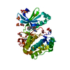
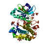
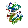
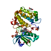
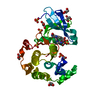
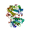
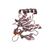
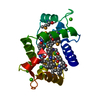
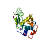
Entry Database : PDB / ID : 2vkiTitle Structure of the PDK1 PH domain K465E mutant 3-PHOPSHOINOSITIDE DEPENDENT PROTEIN KINASE 1 Keywords / / / Function / homology Function Domain/homology Component
/ / / / / / / / / / / / / / / / / / / / / / / / / / / / / / / / / / / / / / / / / / / / / / / / / / / / / / / / / / / / / / / / / / / / / / / / / / / / / / / / / / / / / / / / / / / / / / / / / / / / / Biological species HOMO SAPIENS (human)Method / / Resolution : 1.8 Å Authors Komander, D. / Bayascas, J.R. / Deak, M. / Alessi, D.R. / van Aalten, D.M.F. Journal : Mol.Cell.Biol. / Year : 2008Title : Mutation of the Pdk1 Ph Domain Inhibits Protein Kinase B/Akt, Leading to Small Size and Insulin Resistance.Authors: Bayascas, J.R. / Wullschleger, S. / Sakamoto, K. / Garcia-Martinez, J.M. / Clacher, C. / Komander, D. / Van Aalten, D.M.F. / Boini, K.M. / Lang, F. / Lipina, C. / Logie, L. / Sutherland, C. ... Authors : Bayascas, J.R. / Wullschleger, S. / Sakamoto, K. / Garcia-Martinez, J.M. / Clacher, C. / Komander, D. / Van Aalten, D.M.F. / Boini, K.M. / Lang, F. / Lipina, C. / Logie, L. / Sutherland, C. / Chudek, J.A. / Van Diepen, J.A. / Voshol, P.J. / Lucocq, J.M. / Alessi, D.R. History Deposition Dec 19, 2007 Deposition site / Processing site Revision 1.0 May 13, 2008 Provider / Type Revision 1.1 Jul 13, 2011 Group / Refinement description / Version format complianceRevision 1.2 Dec 13, 2023 Group Data collection / Database references ... Data collection / Database references / Other / Refinement description Category chem_comp_atom / chem_comp_bond ... chem_comp_atom / chem_comp_bond / database_2 / pdbx_database_status / pdbx_initial_refinement_model Item / _database_2.pdbx_database_accession / _pdbx_database_status.status_code_sf
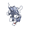
Show all Show less Remark 700 SHEET THE SHEET STRUCTURE OF THIS MOLECULE IS BIFURCATED. IN ORDER TO REPRESENT THIS FEATURE IN ... SHEET THE SHEET STRUCTURE OF THIS MOLECULE IS BIFURCATED. IN ORDER TO REPRESENT THIS FEATURE IN THE SHEET RECORDS BELOW, TWO SHEETS ARE DEFINED.
 Movie
Movie Controller


 Open data
 Basic information
 Components
 Keywords
 Function and homology information
 HOMO SAPIENS (human)
 X-RAY DIFFRACTION /
  Authors
 Citation
 Structure visualization
 Downloads & links
 Other downloads
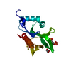


 PDBj
PDBj


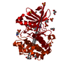
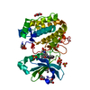
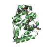
 Assembly


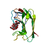
 Mass: 92.094 Da / Num. of mol.: 1 / Source method: obtained synthetically / Formula: C3H8O3
 Mass: 96.063 Da / Num. of mol.: 1 / Source method: obtained synthetically / Formula: SO4
 Mass: 18.015 Da / Num. of mol.: 112 / Source method: isolated from a natural source / Formula: H2O
 Sample preparation
 Processing