 Movie
Movie Controller


+ Open data

- Basic information
| Entry | Database: PDB / ID: 2x10 | ||||||
|---|---|---|---|---|---|---|---|
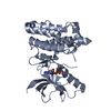
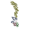
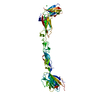
| Title | Crystal structure of the complete EphA2 ectodomain | ||||||
 Components Components | EPHRIN TYPE-A RECEPTOR 2 | ||||||
 Keywords Keywords | RECEPTOR / TRANSFERASE / ANGIOGENESIS / KINASE / CATARACT / APOPTOSIS / GLYCOPROTEIN | ||||||
| Function / homology |  Function and homology information Function and homology informationnotochord cell development / notochord formation / lens fiber cell morphogenesis / blood vessel endothelial cell proliferation involved in sprouting angiogenesis / negative regulation of lymphangiogenesis / axial mesoderm formation / pericyte cell differentiation / cAMP metabolic process / positive regulation of bicellular tight junction assembly / regulation of blood vessel endothelial cell migration ...notochord cell development / notochord formation / lens fiber cell morphogenesis / blood vessel endothelial cell proliferation involved in sprouting angiogenesis / negative regulation of lymphangiogenesis / axial mesoderm formation / pericyte cell differentiation / cAMP metabolic process / positive regulation of bicellular tight junction assembly / regulation of blood vessel endothelial cell migration / ephrin receptor activity / leading edge membrane / negative regulation of chemokine production / post-anal tail morphogenesis / bone remodeling / response to growth factor / tight junction / activation of GTPase activity / regulation of lamellipodium assembly / branching involved in mammary gland duct morphogenesis / EPH-Ephrin signaling / neural tube development / RND1 GTPase cycle / RND2 GTPase cycle / RND3 GTPase cycle / mammary gland epithelial cell proliferation / RHOV GTPase cycle / EPHA-mediated growth cone collapse / growth factor binding / regulation of cell adhesion mediated by integrin / lamellipodium membrane / RHOU GTPase cycle / RHOG GTPase cycle / EPH-ephrin mediated repulsion of cells / ephrin receptor signaling pathway / RAC2 GTPase cycle / RAC3 GTPase cycle / vasculogenesis / transmembrane receptor protein tyrosine kinase activity / regulation of angiogenesis / regulation of ERK1 and ERK2 cascade / negative regulation of angiogenesis / keratinocyte differentiation / RAC1 GTPase cycle / positive regulation of protein localization to plasma membrane / osteoclast differentiation / cell chemotaxis / skeletal system development / molecular function activator activity / protein localization to plasma membrane / cell motility / receptor protein-tyrosine kinase / ruffle membrane / neuron differentiation / osteoblast differentiation / intrinsic apoptotic signaling pathway in response to DNA damage / virus receptor activity / cell migration / lamellipodium / receptor complex / cell adhesion / positive regulation of cell migration / defense response to Gram-positive bacterium / cadherin binding / inflammatory response / focal adhesion / cell surface / ATP binding / plasma membrane Similarity search - Function | ||||||
| Biological species |  Homo sapiens (human) Homo sapiens (human) | ||||||
| Method |  X-RAY DIFFRACTION / SYNCHROTRON / MOLECULAR REPLACEMENT / Resolution: 3 Å X-RAY DIFFRACTION / SYNCHROTRON / MOLECULAR REPLACEMENT / Resolution: 3 Å | ||||||
 Authors Authors | Seiradake, E. / Harlos, K. / Sutton, G. / Aricescu, A.R. / Jones, E.Y. | ||||||
 Citation Citation | Journal: Nat.Struct.Mol.Biol. / Year: 2010 Title: An Extracellular Steric Seeding Mechanism for Eph-Ephrin Signalling Platform Assembly Authors: Seiradake, E. / Harlos, K. / Sutton, G. / Aricescu, A.R. / Jones, E.Y. | ||||||
| History |
|
- Structure visualization
| Structure viewer | Molecule: MolmilJmol/JSmol |
|---|
- Downloads & links
-Download
| PDBx/mmCIF format | 2x10.cif.gz | 203.9 KB | Display | PDBx/mmCIF format |
|---|---|---|---|---|
| PDB format | pdb2x10.ent.gz | 163.8 KB | Display | PDB format |
| PDBx/mmJSON format | 2x10.json.gz | Tree view | PDBx/mmJSON format | |
| Others |  Other downloads Other downloads |
-Validation report
| Summary document | 2x10_validation.pdf.gz | 453.3 KB | Display | wwPDB validaton report |
|---|---|---|---|---|
| Full document | 2x10_full_validation.pdf.gz | 463.5 KB | Display | |
| Data in XML | 2x10_validation.xml.gz | 19.8 KB | Display | |
| Data in CIF | 2x10_validation.cif.gz | 26.5 KB | Display | |
| Arichive directory | https://data.pdbj.org/pub/pdb/validation_reports/x1/2x10ftp://data.pdbj.org/pub/pdb/validation_reports/x1/2x10 | HTTPS FTP |
-Related structure data
| Related structure data |  2x11C  1x5lS  2e7hS  3c8xS C: citing same article ( S: Starting model for refinement |
|---|---|
| Similar structure data |


-Links
 PDBj
PDBj


- Assembly
| Deposited unit | 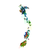
| ||||||||
|---|---|---|---|---|---|---|---|---|---|
| 1 |
| ||||||||
| Unit cell |
|
-Components
| #1: Protein | Mass: 60421.949 Da / Num. of mol.: 1 / Fragment: ECTODOMAIN, RESIDUES 27-534 Source method: isolated from a genetically manipulated source Details: NAG ON ASN407, DI-METHYLATION OF LYSINES / Source: (gene. exp.) Homo sapiens (human) / Plasmid: PHLSEC / Cell line (production host): HEK293 / Production host: Homo sapiens (human) / Variant (production host): GNTI-DEFICIENTReferences: UniProt: P29317, receptor protein-tyrosine kinase | ||
|---|---|---|---|
| #2: Chemical | ChemComp-CL /   Mass: 35.453 Da / Num. of mol.: 1 / Source method: obtained synthetically / Formula: Cl Mass: 35.453 Da / Num. of mol.: 1 / Source method: obtained synthetically / Formula: Cl | ||
| #3: Sugar | ChemComp-NAG /   Type: D-saccharide, beta linking / Mass: 221.208 Da / Num. of mol.: 1 Type: D-saccharide, beta linking / Mass: 221.208 Da / Num. of mol.: 1Source method: isolated from a genetically manipulated source Formula: C8H15NO6 | ||
| Nonpolymer details | N-ACETYLGLUC| Sequence details | INCLUDES FOREIGN N-TERMINAL SIGNAL PEPTIDE AND C-TERMINAL POLY-HIS TAG | |
-Experimental details
-Experiment
| Experiment | Method: X-RAY DIFFRACTION |
|---|
- Sample preparation
| Crystal | Density Matthews: 3.04 Å3/Da / Density % sol: 59.21 % Description: USED PHASES FROM MOLECULAR REPLACEMENT DURING SEARCH FOR SE SITES IN PHASER |
|---|---|
| Crystal grow | Details: 1/3 V/V: PROTEIN, 1/3 V/V: 20 % POLYETHYLENE GLYCOL 6000, 1 M LICL, 0.1 M TRIS PH 8 1/3 V/V: 0.4 M NON-DETERGENT SULFOBETAINE 256 |
-Data collection
| Diffraction | Mean temperature: 100 K |
|---|---|
| Diffraction source | Source: SYNCHROTRON / Site: ESRF  / Beamline: ID23-2 / Wavelength: 0.8726 / Beamline: ID23-2 / Wavelength: 0.8726 |
| Detector | Type: MARRESEARCH / Detector: CCD / Date: Nov 7, 2008 |
| Radiation | Protocol: SINGLE WAVELENGTH / Monochromatic (M) / Laue (L): M / Scattering type: x-ray |
| Radiation wavelength | Wavelength: 0.8726 Å / Relative weight: 1 |
| Reflection | Resolution: 3→91 Å / Num. obs: 23406 / % possible obs: 94.9 % / Observed criterion σ(I): 1 / Redundancy: 1.71 % / Biso Wilson estimate: 53.68 Å2 / Rmerge(I) obs: 0.15 / Net I/σ(I): 6.5 |
| Reflection shell | Resolution: 3→3.1 Å / Redundancy: 1.22 % / Rmerge(I) obs: 0.43 / Mean I/σ(I) obs: 1.6 / % possible all: 82.1 |
- Processing
| Software |
| ||||||||||||||||||||||||||||||||||||||||||||||||||||||||||||||||||||||||||||||||||||||||||||||||||||
|---|---|---|---|---|---|---|---|---|---|---|---|---|---|---|---|---|---|---|---|---|---|---|---|---|---|---|---|---|---|---|---|---|---|---|---|---|---|---|---|---|---|---|---|---|---|---|---|---|---|---|---|---|---|---|---|---|---|---|---|---|---|---|---|---|---|---|---|---|---|---|---|---|---|---|---|---|---|---|---|---|---|---|---|---|---|---|---|---|---|---|---|---|---|---|---|---|---|---|---|---|---|
| Refinement | Method to determine structure: MOLECULAR REPLACEMENT Starting model: PDB ENTRIES 3C8X, 2E7H, 1X5L Resolution: 3→45.917 Å / SU ML: 1.11 / σ(F): 1.98 / Phase error: 33.6 / Stereochemistry target values: ML Details: THE DATA INCLUDES UP TO 2.75 A RESOLUTION, FRIEDEL PAIRS UNMERGED. MERGED PAIRS, UP TO 3A RESOLUTION WERE USED FOR REFINEMENT.
| ||||||||||||||||||||||||||||||||||||||||||||||||||||||||||||||||||||||||||||||||||||||||||||||||||||
| Solvent computation | Shrinkage radii: 0.9 Å / VDW probe radii: 1.11 Å / Solvent model: FLAT BULK SOLVENT MODEL / Bsol: 47.948 Å2 / ksol: 0.253 e/Å3 | ||||||||||||||||||||||||||||||||||||||||||||||||||||||||||||||||||||||||||||||||||||||||||||||||||||
| Displacement parameters |
| ||||||||||||||||||||||||||||||||||||||||||||||||||||||||||||||||||||||||||||||||||||||||||||||||||||
| Refinement step | Cycle: LAST / Resolution: 3→45.917 Å
| ||||||||||||||||||||||||||||||||||||||||||||||||||||||||||||||||||||||||||||||||||||||||||||||||||||
| Refine LS restraints |
| ||||||||||||||||||||||||||||||||||||||||||||||||||||||||||||||||||||||||||||||||||||||||||||||||||||
| LS refinement shell |
| ||||||||||||||||||||||||||||||||||||||||||||||||||||||||||||||||||||||||||||||||||||||||||||||||||||
| Refinement TLS params. | Method: refined / Refine-ID: X-RAY DIFFRACTION
| ||||||||||||||||||||||||||||||||||||||||||||||||||||||||||||||||||||||||||||||||||||||||||||||||||||
| Refinement TLS group |
|
