 Movie
Movie Controller

[English] 日本語
 Yorodumi
- PDB-2x11: Crystal structure of the complete EphA2 ectodomain in complex wit... -
+ Open data

- Basic information
| Entry | Database: PDB / ID: 2x11 | ||||||
|---|---|---|---|---|---|---|---|
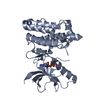
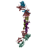
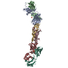
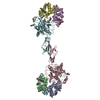
| Title | Crystal structure of the complete EphA2 ectodomain in complex with ephrin A5 receptor binding domain | ||||||
 Components Components |
| ||||||
 Keywords Keywords | RECEPTOR/SIGNALING PROTEIN / RECEPTOR-SIGNALING PROTEIN COMPLEX / DEVELOPMENTAL PROTEIN / SIGNALING PLATFORM / KINASE / TRANSFERASE / NEUROGENESIS / RECEPTOR / CATARACT / APOPTOSIS / ERYTHROPOIETIN-PRODUCING HEPATOCELLULAR CARCINOMA / ANGIOGENESIS / SIGNALING PROTEIN | ||||||
| Function / homology |  Function and homology information Function and homology informationneurotrophin TRKC receptor binding / neurotrophin TRKB receptor binding / positive regulation of collateral sprouting / collateral sprouting / negative regulation of substrate adhesion-dependent cell spreading / notochord cell development / axial mesoderm formation / notochord formation / lens fiber cell morphogenesis / blood vessel endothelial cell proliferation involved in sprouting angiogenesis ...neurotrophin TRKC receptor binding / neurotrophin TRKB receptor binding / positive regulation of collateral sprouting / collateral sprouting / negative regulation of substrate adhesion-dependent cell spreading / notochord cell development / axial mesoderm formation / notochord formation / lens fiber cell morphogenesis / blood vessel endothelial cell proliferation involved in sprouting angiogenesis / negative regulation of lymphangiogenesis / synaptic membrane adhesion / central nervous system neuron differentiation / branching involved in mammary gland duct morphogenesis / cAMP metabolic process / regulation of blood vessel endothelial cell migration / pericyte cell differentiation / cellular response to follicle-stimulating hormone stimulus / leading edge membrane / regulation of insulin secretion involved in cellular response to glucose stimulus / negative regulation of chemokine production / post-anal tail morphogenesis / transmembrane receptor protein tyrosine kinase activator activity / ephrin receptor activity / bone remodeling / response to growth factor / activation of GTPase activity / neurotrophin TRKA receptor binding / chemorepellent activity / positive regulation of peptidyl-tyrosine phosphorylation / positive regulation of bicellular tight junction assembly / regulation of cell morphogenesis / regulation of GTPase activity / regulation of lamellipodium assembly / positive regulation of synapse assembly / negative regulation of cell adhesion mediated by integrin / neural tube development / EPH-Ephrin signaling / retinal ganglion cell axon guidance / mammary gland epithelial cell proliferation / RND1 GTPase cycle / RND2 GTPase cycle / RND3 GTPase cycle / regulation of focal adhesion assembly / tight junction / RHOV GTPase cycle / EPHA-mediated growth cone collapse / growth factor binding / regulation of cell-cell adhesion / RHOU GTPase cycle / lamellipodium membrane / basement membrane / RHOG GTPase cycle / keratinocyte differentiation / vasculogenesis / EPH-ephrin mediated repulsion of cells / regulation of angiogenesis / RAC3 GTPase cycle / RAC2 GTPase cycle / ephrin receptor signaling pathway / regulation of ERK1 and ERK2 cascade / osteoclast differentiation / ephrin receptor binding / RAC1 GTPase cycle / transmembrane receptor protein tyrosine kinase activity / negative regulation of angiogenesis / cell surface receptor protein tyrosine kinase signaling pathway / axon guidance / molecular function activator activity / skeletal system development / cellular response to forskolin / protein localization to plasma membrane / regulation of actin cytoskeleton organization / regulation of microtubule cytoskeleton organization / positive regulation of protein localization to plasma membrane / cell motility / cell chemotaxis / adherens junction / receptor protein-tyrosine kinase / intrinsic apoptotic signaling pathway in response to DNA damage / caveola / GABA-ergic synapse / ruffle membrane / osteoblast differentiation / cell migration / nervous system development / lamellipodium / virus receptor activity / angiogenesis / positive regulation of phosphatidylinositol 3-kinase/protein kinase B signal transduction / signaling receptor complex / cell adhesion / defense response to Gram-positive bacterium / positive regulation of cell migration / cadherin binding / inflammatory response / receptor ligand activity / external side of plasma membrane / focal adhesion / cell surface Similarity search - Function | ||||||
| Biological species |  Homo sapiens (human) Homo sapiens (human) | ||||||
| Method |  X-RAY DIFFRACTION / SYNCHROTRON / MOLECULAR REPLACEMENT / Resolution: 4.83 Å X-RAY DIFFRACTION / SYNCHROTRON / MOLECULAR REPLACEMENT / Resolution: 4.83 Å | ||||||
 Authors Authors | Seiradake, E. / Harlos, K. / Sutton, G. / Aricescu, A.R. / Jones, E.Y. | ||||||
 Citation Citation | Journal: Nat.Struct.Mol.Biol. / Year: 2010 Title: An Extracellular Steric Seeding Mechanism for Eph-Ephrin Signalling Platform Assembly Authors: Seiradake, E. / Harlos, K. / Sutton, G. / Aricescu, A.R. / Jones, E.Y. | ||||||
| History |
|
- Structure visualization
| Structure viewer | Molecule: MolmilJmol/JSmol |
|---|
- Downloads & links
-Download
| PDBx/mmCIF format | 2x11.cif.gz | 260.5 KB | Display | PDBx/mmCIF format |
|---|---|---|---|---|
| PDB format | pdb2x11.ent.gz | 213.6 KB | Display | PDB format |
| PDBx/mmJSON format | 2x11.json.gz | Tree view | PDBx/mmJSON format | |
| Others |  Other downloads Other downloads |
-Validation report
| Arichive directory | https://data.pdbj.org/pub/pdb/validation_reports/x1/2x11ftp://data.pdbj.org/pub/pdb/validation_reports/x1/2x11 | HTTPS FTP |
|---|
-Related structure data
| Related structure data |  2x10C  1shwS  3czuS  3fl7S C: citing same article ( S: Starting model for refinement |
|---|---|
| Similar structure data |


-Links
 PDBj
PDBj


- Assembly
| Deposited unit | 
| ||||||||
|---|---|---|---|---|---|---|---|---|---|
| 1 |
| ||||||||
| Unit cell |
|
-Components
| #1: Protein | Mass: 59859.215 Da / Num. of mol.: 1 / Fragment: ECTODOMAIN, RESIDUES 27-534 Source method: isolated from a genetically manipulated source Details: NAG ON ASN407, DI-METHYLATION OF LYSINES / Source: (gene. exp.) Homo sapiens (human) / Plasmid: PHLSEC / Cell line (production host): HEK293 / Production host: Homo sapiens (human) / Variant (production host): GNTI-DEFICIENTReferences: UniProt: P29317, receptor protein-tyrosine kinase |
|---|---|
| #2: Protein | Mass: 20402.324 Da / Num. of mol.: 1 / Fragment: RECEPTOR BINDING DOMAIN, RESIDUES 26-166 Source method: isolated from a genetically manipulated source Source: (gene. exp.) Homo sapiens (human) / Plasmid: PHLSEC / Cell line (production host): HEK293 / Production host: Homo sapiens (human) / Variant (production host): GNTI-DEFICIENT / References: UniProt: P52803 |
| Has protein modification | Y |
| Sequence details | CONTAINS FOREIGN SIGNAL PEPTIDE AND POLY-HIS TAG |
-Experimental details
-Experiment
| Experiment | Method: X-RAY DIFFRACTION / Number of used crystals: 1 |
|---|
- Sample preparation
| Crystal | Density Matthews: 3.61 Å3/Da / Density % sol: 66 % / Description: NONE |
|---|---|
| Crystal grow | Details: TWO PARTS OF PROTEIN SOLUTION WERE MIXED WITH ONE PART WATER, ONE PART RESERVOIR SOLUTION (8 % POLYETHYLENE GLYCOL 6000, 0.8 M LICL, 0.08 M CITRATE PH 5) AND ONE PART 1 % POLYVINYLPYRROLIDONE K15. |
-Data collection
| Diffraction | Mean temperature: 100 K |
|---|---|
| Diffraction source | Source: SYNCHROTRON / Site: ESRF  / Beamline: ID14-2 / Wavelength: 0.933 / Beamline: ID14-2 / Wavelength: 0.933 |
| Detector | Type: ADSC QUANTUM 4 / Detector: CCD / Date: Dec 13, 2008 |
| Radiation | Protocol: SINGLE WAVELENGTH / Monochromatic (M) / Laue (L): M / Scattering type: x-ray |
| Radiation wavelength | Wavelength: 0.933 Å / Relative weight: 1 |
| Reflection | Resolution: 4.8→173 Å / Num. obs: 6016 / % possible obs: 98.2 % / Observed criterion σ(I): 1 / Redundancy: 6.2 % / Biso Wilson estimate: 166.11 Å2 / Rmerge(I) obs: 0.11 / Net I/σ(I): 12.95 |
| Reflection shell | Resolution: 4.8→4.9 Å / Redundancy: 6.2 % / Rmerge(I) obs: 0.74 / Mean I/σ(I) obs: 2.8 / % possible all: 75.7 |
- Processing
| Software |
| ||||||||||||||||||||||||
|---|---|---|---|---|---|---|---|---|---|---|---|---|---|---|---|---|---|---|---|---|---|---|---|---|---|
| Refinement | Method to determine structure: MOLECULAR REPLACEMENT Starting model: PDB ENTRIES 3CZU, 3FL7, 1SHW Resolution: 4.83→40.482 Å / σ(F): 0.03 / Phase error: 34 / Stereochemistry target values: ML Details: DUE TO LOW RESOLUTION, THE REFINEMENT PROTOCOL WAS LIMITED TO THE FOLLOWING THREE STEPS AFTER MOLECULAR REPLACEMENT. 1. RIGID BODY REFINEMENT OF INDIVIDUAL DOMAINS 2. TLS REFINEMENT OF ...Details: DUE TO LOW RESOLUTION, THE REFINEMENT PROTOCOL WAS LIMITED TO THE FOLLOWING THREE STEPS AFTER MOLECULAR REPLACEMENT. 1. RIGID BODY REFINEMENT OF INDIVIDUAL DOMAINS 2. TLS REFINEMENT OF SELECTED PORTIONS 3. STRUCTURE REGULARIZATION TO AVOID MINOR CLASHES. WE PERFORMED NO INDIVIDUAL ATOM REFINEMENT.
| ||||||||||||||||||||||||
| Solvent computation | Shrinkage radii: 0.9 Å / VDW probe radii: 1.11 Å / Solvent model: FLAT BULK SOLVENT MODEL / Bsol: 99 Å2 / ksol: 0.298 e/Å3 | ||||||||||||||||||||||||
| Displacement parameters |
| ||||||||||||||||||||||||
| Refinement step | Cycle: LAST / Resolution: 4.83→40.482 Å
| ||||||||||||||||||||||||
| Refine LS restraints |
| ||||||||||||||||||||||||
| LS refinement shell |
|
