 Movie
Movie Controller


+ Open data

- Basic information
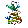
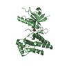
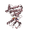
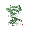
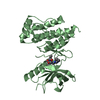
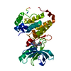
| Entry | Database: PDB / ID: 1mqb | ||||||
|---|---|---|---|---|---|---|---|
| Title | Crystal Structure of Ephrin A2 (ephA2) Receptor Protein Kinase | ||||||
 Components Components | Ephrin type-A receptor 2 | ||||||
 Keywords Keywords | TRANSFERASE / Tyrosine Protein Kinase | ||||||
| Function / homology |  Function and homology information Function and homology informationnotochord cell development / notochord formation / lens fiber cell morphogenesis / blood vessel endothelial cell proliferation involved in sprouting angiogenesis / negative regulation of lymphangiogenesis / axial mesoderm formation / cAMP metabolic process / regulation of blood vessel endothelial cell migration / pericyte cell differentiation / leading edge membrane ...notochord cell development / notochord formation / lens fiber cell morphogenesis / blood vessel endothelial cell proliferation involved in sprouting angiogenesis / negative regulation of lymphangiogenesis / axial mesoderm formation / cAMP metabolic process / regulation of blood vessel endothelial cell migration / pericyte cell differentiation / leading edge membrane / negative regulation of chemokine production / ephrin receptor activity / activation of GTPase activity / response to growth factor / post-anal tail morphogenesis / bone remodeling / positive regulation of bicellular tight junction assembly / regulation of lamellipodium assembly / negative regulation of cell adhesion mediated by integrin / branching involved in mammary gland duct morphogenesis / central nervous system neuron differentiation / EPH-Ephrin signaling / RND1 GTPase cycle / RND2 GTPase cycle / RND3 GTPase cycle / neural tube development / mammary gland epithelial cell proliferation / tight junction / RHOV GTPase cycle / EPHA-mediated growth cone collapse / growth factor binding / RHOU GTPase cycle / lamellipodium membrane / RHOG GTPase cycle / EPH-ephrin mediated repulsion of cells / RAC3 GTPase cycle / regulation of angiogenesis / RAC2 GTPase cycle / ephrin receptor signaling pathway / vasculogenesis / regulation of ERK1 and ERK2 cascade / keratinocyte differentiation / RAC1 GTPase cycle / transmembrane receptor protein tyrosine kinase activity / osteoclast differentiation / negative regulation of angiogenesis / cell surface receptor protein tyrosine kinase signaling pathway / molecular function activator activity / protein localization to plasma membrane / positive regulation of protein localization to plasma membrane / cell chemotaxis / skeletal system development / cell motility / receptor protein-tyrosine kinase / intrinsic apoptotic signaling pathway in response to DNA damage / ruffle membrane / osteoblast differentiation / cell migration / lamellipodium / virus receptor activity / angiogenesis / cell adhesion / signaling receptor complex / defense response to Gram-positive bacterium / positive regulation of cell migration / cadherin binding / inflammatory response / focal adhesion / cell surface / ATP binding / plasma membrane Similarity search - Function | ||||||
| Biological species |  Homo sapiens (human) Homo sapiens (human) | ||||||
| Method |  X-RAY DIFFRACTION / SYNCHROTRON / MOLECULAR REPLACEMENT / Resolution: 2.3 Å X-RAY DIFFRACTION / SYNCHROTRON / MOLECULAR REPLACEMENT / Resolution: 2.3 Å | ||||||
 Authors Authors | Nowakowski, J. / Cronin, C.N. / McRee, D.E. / Knuth, M.W. / Nelson, C. / Pavletich, N. / Rogers, J. / Sang, B.C. / Scheibe, D.N. / Swanson, R.V. / Thompson, D.A. | ||||||
 Citation Citation | Journal: Structure / Year: 2003 Title: Structures of the Cancer Related Aurora-A, FAK and EphA2 Protein Kinases from Nanovolume Crystallography Authors: Nowakowski, J. / Cronin, C.N. / McRee, D.E. / Knuth, M.W. / Nelson, C. / Pavletich, N. / Rogers, J. / Sang, B.C. / Scheibe, D.N. / Swanson, R.V. / Thompson, D.A. | ||||||
| History |
|
- Structure visualization
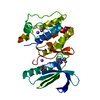
| Structure viewer | Molecule: MolmilJmol/JSmol |
|---|
- Downloads & links
-Download
| PDBx/mmCIF format | 1mqb.cif.gz | 121.1 KB | Display | PDBx/mmCIF format |
|---|---|---|---|---|
| PDB format | pdb1mqb.ent.gz | 93.4 KB | Display | PDB format |
| PDBx/mmJSON format | 1mqb.json.gz | Tree view | PDBx/mmJSON format | |
| Others |  Other downloads Other downloads |
-Validation report
| Arichive directory | https://data.pdbj.org/pub/pdb/validation_reports/mq/1mqbftp://data.pdbj.org/pub/pdb/validation_reports/mq/1mqb | HTTPS FTP |
|---|
-Related structure data
| Related structure data | 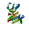 1mp8C 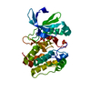 1mq4C  1jpaS C: citing same article ( S: Starting model for refinement |
|---|---|
| Similar structure data |


-Links
 PDBj
PDBj


- Assembly
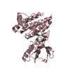
| Deposited unit | 
| ||||||||
|---|---|---|---|---|---|---|---|---|---|
| 1 | 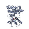
| ||||||||
| 2 | 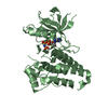
| ||||||||
| Unit cell |
|
-Components
| #1: Protein | Mass: 37783.410 Da / Num. of mol.: 2 / Fragment: Kinase Domain Source method: isolated from a genetically manipulated source Source: (gene. exp.) Homo sapiens (human) / Gene: EphA2 / Cell line (production host): Hi5 / Production host:   Spodoptera frugiperda (fall armyworm) / References: UniProt: P29317, EC: 2.7.1.112 Spodoptera frugiperda (fall armyworm) / References: UniProt: P29317, EC: 2.7.1.112#2: Chemical |   Mass: 506.196 Da / Num. of mol.: 2 / Source method: obtained synthetically / Formula: C10H17N6O12P3 / Comment: AMP-PNP, energy-carrying molecule analogue*YM Mass: 506.196 Da / Num. of mol.: 2 / Source method: obtained synthetically / Formula: C10H17N6O12P3 / Comment: AMP-PNP, energy-carrying molecule analogue*YM#3: Water | ChemComp-HOH / |  Mass: 18.015 Da / Num. of mol.: 66 / Source method: isolated from a natural source / Formula: H2O Mass: 18.015 Da / Num. of mol.: 66 / Source method: isolated from a natural source / Formula: H2O |
|---|
-Experimental details
-Experiment
| Experiment | Method: X-RAY DIFFRACTION / Number of used crystals: 1 |
|---|
- Sample preparation
| Crystal | Density Matthews: 2.4 Å3/Da / Density % sol: 48.75 % | |||||||||||||||||||||||||||||||||||||||||||||||||||||||||||||||
|---|---|---|---|---|---|---|---|---|---|---|---|---|---|---|---|---|---|---|---|---|---|---|---|---|---|---|---|---|---|---|---|---|---|---|---|---|---|---|---|---|---|---|---|---|---|---|---|---|---|---|---|---|---|---|---|---|---|---|---|---|---|---|---|---|
| Crystal grow | Temperature: 293 K / Method: vapor diffusion, sitting drop / pH: 7.5 Details: Ethylene Glycol, PEG 10K, HEPES, pH 7.5, VAPOR DIFFUSION, SITTING DROP, temperature 293K | |||||||||||||||||||||||||||||||||||||||||||||||||||||||||||||||
| Crystal grow | *PLUS Temperature: 4 ℃ / pH: 7.6 / Method: vapor diffusion, sitting drop | |||||||||||||||||||||||||||||||||||||||||||||||||||||||||||||||
| Components of the solutions | *PLUS
|
-Data collection
| Diffraction | Mean temperature: 100 K |
|---|---|
| Diffraction source | Source: SYNCHROTRON / Site: ALS  / Beamline: 5.0.3 / Wavelength: 1 Å / Beamline: 5.0.3 / Wavelength: 1 Å |
| Detector | Type: ADSC QUANTUM 4 / Detector: CCD / Date: Dec 12, 2001 / Details: synchrotron |
| Radiation | Monochromator: crystal / Protocol: SINGLE WAVELENGTH / Monochromatic (M) / Laue (L): M / Scattering type: x-ray |
| Radiation wavelength | Wavelength: 1 Å / Relative weight: 1 |
| Reflection | Resolution: 2.3→40 Å / Num. all: 36207 / Num. obs: 35483 / % possible obs: 98 % / Observed criterion σ(F): 2 / Observed criterion σ(I): 2 / Redundancy: 4.1 % / Biso Wilson estimate: 44.66 Å2 / Rmerge(I) obs: 0.057 / Rsym value: 0.057 / Net I/σ(I): 11.5 |
| Reflection shell | Resolution: 2.3→2.36 Å / Redundancy: 3.8 % / Rmerge(I) obs: 0.485 / Mean I/σ(I) obs: 2 / Num. unique all: 1662 / Rsym value: 0.485 / % possible all: 0.78 |
| Reflection | *PLUS Lowest resolution: 63 Å / % possible obs: 95 % |
| Reflection shell | *PLUS % possible obs: 78 % |
- Processing
| Software |
| ||||||||||||||||||||||||||||||||||||||||||||||||||||||||||||||||||||||||||||||||||||||||||||||||||||
|---|---|---|---|---|---|---|---|---|---|---|---|---|---|---|---|---|---|---|---|---|---|---|---|---|---|---|---|---|---|---|---|---|---|---|---|---|---|---|---|---|---|---|---|---|---|---|---|---|---|---|---|---|---|---|---|---|---|---|---|---|---|---|---|---|---|---|---|---|---|---|---|---|---|---|---|---|---|---|---|---|---|---|---|---|---|---|---|---|---|---|---|---|---|---|---|---|---|---|---|---|---|
| Refinement | Method to determine structure: MOLECULAR REPLACEMENT Starting model: 1JPA Resolution: 2.3→40 Å / Cor.coef. Fo:Fc: 0.927 / Cor.coef. Fo:Fc free: 0.885 / SU B: 8.733 / SU ML: 0.208 / Cross valid method: THROUGHOUT / σ(F): 0 / σ(I): 0 / ESU R: 0.324 / ESU R Free: 0.267 / Stereochemistry target values: MAXIMUM LIKELIHOOD / Details: HYDROGENS HAVE BEEN ADDED IN THE RIDING POSITIONS
| ||||||||||||||||||||||||||||||||||||||||||||||||||||||||||||||||||||||||||||||||||||||||||||||||||||
| Solvent computation | Ion probe radii: 0.8 Å / Shrinkage radii: 0.8 Å / VDW probe radii: 1.4 Å / Solvent model: BABINET MODEL WITH MASK | ||||||||||||||||||||||||||||||||||||||||||||||||||||||||||||||||||||||||||||||||||||||||||||||||||||
| Displacement parameters | Biso mean: 44.668 Å2
| ||||||||||||||||||||||||||||||||||||||||||||||||||||||||||||||||||||||||||||||||||||||||||||||||||||
| Refinement step | Cycle: LAST / Resolution: 2.3→40 Å
| ||||||||||||||||||||||||||||||||||||||||||||||||||||||||||||||||||||||||||||||||||||||||||||||||||||
| Refine LS restraints |
| ||||||||||||||||||||||||||||||||||||||||||||||||||||||||||||||||||||||||||||||||||||||||||||||||||||
| LS refinement shell | Resolution: 2.3→2.36 Å / Total num. of bins used: 20 /
| ||||||||||||||||||||||||||||||||||||||||||||||||||||||||||||||||||||||||||||||||||||||||||||||||||||
| Refinement | *PLUS Highest resolution: 2.3 Å / Lowest resolution: 63 Å / Num. reflection obs: 30484 / % reflection Rfree: 5 % / Rfactor Rfree: 0.289 / Rfactor Rwork: 0.235 | ||||||||||||||||||||||||||||||||||||||||||||||||||||||||||||||||||||||||||||||||||||||||||||||||||||
| Solvent computation | *PLUS | ||||||||||||||||||||||||||||||||||||||||||||||||||||||||||||||||||||||||||||||||||||||||||||||||||||
| Displacement parameters | *PLUS | ||||||||||||||||||||||||||||||||||||||||||||||||||||||||||||||||||||||||||||||||||||||||||||||||||||
| Refine LS restraints | *PLUS
|
