positive regulation of adaptive immune memory response / positive regulation of protein catabolic process in the vacuole / CD4-positive, alpha-beta T cell costimulation / positive regulation of B cell receptor signaling pathway / osteoclast fusion / myoblast fusion involved in skeletal muscle regeneration / positive regulation of T cell activation via T cell receptor contact with antigen bound to MHC molecule on antigen presenting cell / positive regulation of inflammatory response to antigenic stimulus / regulation of macrophage migration / macrophage fusion ...positive regulation of adaptive immune memory response / positive regulation of protein catabolic process in the vacuole / CD4-positive, alpha-beta T cell costimulation / positive regulation of B cell receptor signaling pathway / osteoclast fusion / myoblast fusion involved in skeletal muscle regeneration / positive regulation of T cell activation via T cell receptor contact with antigen bound to MHC molecule on antigen presenting cell / positive regulation of inflammatory response to antigenic stimulus / regulation of macrophage migration / macrophage fusion / immunological synapse formation / tetraspanin-enriched microdomain / positive regulation of T-helper 2 cell cytokine production / transferrin receptor binding / humoral immune response mediated by circulating immunoglobulin / positive regulation of protein exit from endoplasmic reticulum / protein localization to lysosome / MHC class II protein binding / positive regulation of CD4-positive, alpha-beta T cell proliferation / positive regulation of T cell receptor signaling pathway / cholesterol binding / immunological synapse / cellular response to low-density lipoprotein particle stimulus / positive regulation of receptor clustering / positive regulation of B cell proliferation / Regulation of Complement cascade / basal plasma membrane / protein localization to plasma membrane / regulation of protein stability / receptor internalization / integrin binding / Immunoregulatory interactions between a Lymphoid and a non-Lymphoid cell / MHC class II protein complex binding / virus receptor activity / vesicle / basolateral plasma membrane / positive regulation of MAPK cascade / focal adhesion / positive regulation of transcription by RNA polymerase II / extracellular exosome / membrane / plasma membrane / cytosol Similarity search - Function
Protocol: SINGLE WAVELENGTH / Monochromatic (M) / Laue (L): M / Scattering type: x-ray
Radiation wavelength
Wavelength: 0.97946 Å / Relative weight: 1
Reflection
Resolution: 2.8→50 Å / Num. obs: 30682 / % possible obs: 94 % / Redundancy: 3.4 % / Rsym value: 0.125 / Net I/σ(I): 7.1
Reflection shell
Resolution: 2.8→2.93 Å / Redundancy: 1.9 % / Rmerge(I) obs: 0.442 / Mean I/σ(I) obs: 1.4 / % possible all: 63.7
-
Processing
Software
Name
Version
Classification
REFMAC
5.2.0019
refinement
SCALEPACK
datascaling
DENZO
datareduction
PHASER
phasing
Refinement
Method to determine structure: MOLECULAR REPLACEMENT / Resolution: 2.8→47.25 Å / Cor.coef. Fo:Fc: 0.889 / Cor.coef. Fo:Fc free: 0.83 / SU B: 17.241 / SU ML: 0.332 / Cross valid method: THROUGHOUT / ESU R Free: 0.442 / Stereochemistry target values: MAXIMUM LIKELIHOOD / Details: HYDROGENS HAVE BEEN ADDED IN THE RIDING POSITIONS
Rfactor
Num. reflection
% reflection
Selection details
Rfree
0.29066
1583
5.2 %
RANDOM
Rwork
0.23947
-
-
-
obs
0.24213
29086
93.98 %
-
Solvent computation
Ion probe radii: 0.8 Å / Shrinkage radii: 0.8 Å / VDW probe radii: 1.2 Å / Solvent model: MASK
 Movie
Movie Controller

 Yorodumi
 Open data
 Basic information
 Components
 Keywords
 Function and homology information
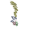
 Homo sapiens (human)
 X-RAY DIFFRACTION /
  Authors
 Citation
 Structure visualization
 Downloads & links
 Other downloads


 PDBj
PDBj

 Assembly

 Mass: 18.015 Da / Num. of mol.: 4 / Source method: isolated from a natural source / Formula: H2O
 Sample preparation
 / Beamline: BL7-1 / Wavelength: 0.97946 Å
 Processing