 Movie
Movie Controller


+ Open data

- Basic information
| Entry | Database: PDB / ID: 2iwp | ||||||
|---|---|---|---|---|---|---|---|
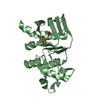
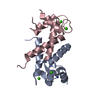
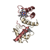
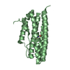
| Title | 12th PDZ domain of Multiple PDZ Domain Protein MPDZ | ||||||
 Components Components | MULTIPLE PDZ DOMAIN PROTEIN | ||||||
 Keywords Keywords | SIGNALING PROTEIN / SGC / PDZ / MPDZ / MUPP1 / MUPP-1 / MEMBRANE / PDZ DOMAIN / HOST- VIRUS INTERACTION / STRUCTURAL GENOMICS CONSORTIUM / SYNAPTOSOME / TIGHT JUNCTION | ||||||
| Function / homology |  Function and homology information Function and homology informationtight junction assembly / apicolateral plasma membrane / bicellular tight junction / apical part of cell / apical plasma membrane / postsynaptic density / dendrite / plasma membrane / cytoplasm Similarity search - Function | ||||||
| Biological species |  HOMO SAPIENS (human) HOMO SAPIENS (human) | ||||||
| Method |  X-RAY DIFFRACTION / SYNCHROTRON / MOLECULAR REPLACEMENT / Resolution: 2.15 Å X-RAY DIFFRACTION / SYNCHROTRON / MOLECULAR REPLACEMENT / Resolution: 2.15 Å | ||||||
 Authors Authors | Elkins, J.M. / Yang, X. / Gileadi, C. / Schoch, G. / Johansson, C. / Savitsky, P. / Berridge, G. / Smee, C.E.A. / Turnbull, A. / Pike, A. ...Elkins, J.M. / Yang, X. / Gileadi, C. / Schoch, G. / Johansson, C. / Savitsky, P. / Berridge, G. / Smee, C.E.A. / Turnbull, A. / Pike, A. / Papagrigoriou, E. / Sundstrom, M. / Edwards, A. / Arrowsmith, C. / Weigelt, J. / Doyle, D.A. | ||||||
 Citation Citation | Journal: Protein Sci. / Year: 2007 Title: Structure of Pick1 and Other Pdz Domains Obtained with the Help of Self-Binding C-Terminal Extension. Authors: Elkins, J.M. / Papagrigoriou, E. / Berridge, G. / Yang, X. / Phillips, C. / Gileadi, C. / Savitsky, P. / Doyle, D.A. | ||||||
| History |
|
- Structure visualization
| Structure viewer | Molecule: MolmilJmol/JSmol |
|---|
- Downloads & links
-Download
| PDBx/mmCIF format | 2iwp.cif.gz | 48.6 KB | Display | PDBx/mmCIF format |
|---|---|---|---|---|
| PDB format | pdb2iwp.ent.gz | 34.3 KB | Display | PDB format |
| PDBx/mmJSON format | 2iwp.json.gz | Tree view | PDBx/mmJSON format | |
| Others |  Other downloads Other downloads |
-Validation report
| Arichive directory | https://data.pdbj.org/pub/pdb/validation_reports/iw/2iwpftp://data.pdbj.org/pub/pdb/validation_reports/iw/2iwp | HTTPS FTP |
|---|
-Related structure data
| Related structure data |  2bygC  2fcfC  2fneSC  2gzvC  2he2C  2he4C  2i1nC  2iwnC  2iwoC  2iwqC C: citing same article ( S: Starting model for refinement |
|---|---|
| Similar structure data |


-Links
 PDBj
PDBj- Assembly
| Deposited unit | 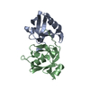
| ||||||||||||||||||||||||||||||||||||||||||||||||||||||||||||||||||||||||||
|---|---|---|---|---|---|---|---|---|---|---|---|---|---|---|---|---|---|---|---|---|---|---|---|---|---|---|---|---|---|---|---|---|---|---|---|---|---|---|---|---|---|---|---|---|---|---|---|---|---|---|---|---|---|---|---|---|---|---|---|---|---|---|---|---|---|---|---|---|---|---|---|---|---|---|---|
| 1 |
| ||||||||||||||||||||||||||||||||||||||||||||||||||||||||||||||||||||||||||
| Unit cell |
| ||||||||||||||||||||||||||||||||||||||||||||||||||||||||||||||||||||||||||
| Noncrystallographic symmetry (NCS) | NCS domain:
NCS domain segments:
NCS ensembles :
NCS oper: (Code: given Matrix: (-0.99871, -0.03148, -0.03987), Vector: Details | BIOLOGICAL ASSEMBLY: MONOMERIC | |
-Components
| #1: Protein | Mass: 12550.251 Da / Num. of mol.: 2 / Fragment: 12TH PDZ DOMAIN, RESIDUES 1831-1923 Source method: isolated from a genetically manipulated source Source: (gene. exp.) HOMO SAPIENS (human) / Plasmid: PNIC28-BSA4 / Production host:  #2: Water | ChemComp-HOH / |  Mass: 18.015 Da / Num. of mol.: 24 / Source method: isolated from a natural source / Formula: H2O Mass: 18.015 Da / Num. of mol.: 24 / Source method: isolated from a natural source / Formula: H2O |
|---|
-Experimental details
-Experiment
| Experiment | Method: X-RAY DIFFRACTION / Number of used crystals: 1 |
|---|
- Sample preparation
| Crystal | Density Matthews: 2.46 Å3/Da / Density % sol: 50.1 % / Description: NONE |
|---|---|
| Crystal grow | pH: 4.5 / Details: 50% PEG 300, 0.2M LI2SO4, 0.1M ACETATE PH 4.5 |
-Data collection
| Diffraction | Mean temperature: 100 K |
|---|---|
| Diffraction source | Source: SYNCHROTRON / Site: SLS  / Beamline: X10SA / Wavelength: 0.976 / Beamline: X10SA / Wavelength: 0.976 |
| Detector | Type: MARRESEARCH / Detector: CCD / Date: Jun 16, 2006 |
| Radiation | Protocol: SINGLE WAVELENGTH / Monochromatic (M) / Laue (L): M / Scattering type: x-ray |
| Radiation wavelength | Wavelength: 0.976 Å / Relative weight: 1 |
| Reflection | Resolution: 2.15→57.83 Å / Num. obs: 13731 / % possible obs: 99.4 % / Observed criterion σ(I): 0 / Redundancy: 3.7 % / Rmerge(I) obs: 0.09 / Net I/σ(I): 11 |
| Reflection shell | Resolution: 2.15→2.27 Å / Redundancy: 3.7 % / Rmerge(I) obs: 0.47 / Mean I/σ(I) obs: 2.5 / % possible all: 100 |
- Processing
| Software |
| ||||||||||||||||||||||||||||||||||||||||||||||||||||||||||||||||||||||||||||||||||||||||||||||||||||||||||||||||||||||||||||||||||||||||||||||||||||||||||||||||||||||||||||||||||||||
|---|---|---|---|---|---|---|---|---|---|---|---|---|---|---|---|---|---|---|---|---|---|---|---|---|---|---|---|---|---|---|---|---|---|---|---|---|---|---|---|---|---|---|---|---|---|---|---|---|---|---|---|---|---|---|---|---|---|---|---|---|---|---|---|---|---|---|---|---|---|---|---|---|---|---|---|---|---|---|---|---|---|---|---|---|---|---|---|---|---|---|---|---|---|---|---|---|---|---|---|---|---|---|---|---|---|---|---|---|---|---|---|---|---|---|---|---|---|---|---|---|---|---|---|---|---|---|---|---|---|---|---|---|---|---|---|---|---|---|---|---|---|---|---|---|---|---|---|---|---|---|---|---|---|---|---|---|---|---|---|---|---|---|---|---|---|---|---|---|---|---|---|---|---|---|---|---|---|---|---|---|---|---|---|
| Refinement | Method to determine structure: MOLECULAR REPLACEMENT Starting model: PDB ENTRY 2FNE Resolution: 2.15→57.83 Å / Cor.coef. Fo:Fc: 0.947 / Cor.coef. Fo:Fc free: 0.916 / SU B: 12.001 / SU ML: 0.156 / TLS residual ADP flag: LIKELY RESIDUAL / Cross valid method: THROUGHOUT / ESU R: 0.21 / ESU R Free: 0.194 / Stereochemistry target values: MAXIMUM LIKELIHOOD / Details: HYDROGENS HAVE BEEN ADDED IN THE RIDING POSITIONS.
| ||||||||||||||||||||||||||||||||||||||||||||||||||||||||||||||||||||||||||||||||||||||||||||||||||||||||||||||||||||||||||||||||||||||||||||||||||||||||||||||||||||||||||||||||||||||
| Solvent computation | Ion probe radii: 0.8 Å / Shrinkage radii: 0.8 Å / VDW probe radii: 1.4 Å / Solvent model: MASK | ||||||||||||||||||||||||||||||||||||||||||||||||||||||||||||||||||||||||||||||||||||||||||||||||||||||||||||||||||||||||||||||||||||||||||||||||||||||||||||||||||||||||||||||||||||||
| Displacement parameters | Biso mean: 44.16 Å2
| ||||||||||||||||||||||||||||||||||||||||||||||||||||||||||||||||||||||||||||||||||||||||||||||||||||||||||||||||||||||||||||||||||||||||||||||||||||||||||||||||||||||||||||||||||||||
| Refinement step | Cycle: LAST / Resolution: 2.15→57.83 Å
| ||||||||||||||||||||||||||||||||||||||||||||||||||||||||||||||||||||||||||||||||||||||||||||||||||||||||||||||||||||||||||||||||||||||||||||||||||||||||||||||||||||||||||||||||||||||
| Refine LS restraints |
|