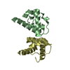
 Movie
Movie Controller


+ Open data

- Basic information
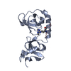
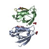
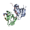
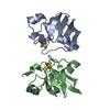
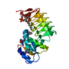
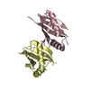
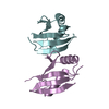
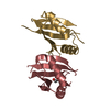
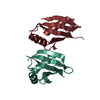
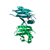
| Entry | Database: PDB / ID: 4mjs | ||||||
|---|---|---|---|---|---|---|---|
| Title | crystal structure of a PB1 complex | ||||||
 Components Components |
| ||||||
 Keywords Keywords | TRANSFERASE/PROTEIN BINDING / PB1 domain / PB1 heterodimer and protein interaction / TRANSFERASE-PROTEIN BINDING complex | ||||||
| Function / homology |  Function and homology information Function and homology informationTGF-beta receptor signaling in EMT (epithelial to mesenchymal transition) / VEGFR2 mediated cell proliferation / calcium,diacylglycerol-dependent serine/threonine kinase activity / protein kinase C signaling / diacylglycerol-dependent, calcium-independent serine/threonine kinase activity / protein localization to perinuclear region of cytoplasm / brown fat cell proliferation / protein targeting to vacuole involved in autophagy / regulation of Ras protein signal transduction / positive regulation of protein transport ...TGF-beta receptor signaling in EMT (epithelial to mesenchymal transition) / VEGFR2 mediated cell proliferation / calcium,diacylglycerol-dependent serine/threonine kinase activity / protein kinase C signaling / diacylglycerol-dependent, calcium-independent serine/threonine kinase activity / protein localization to perinuclear region of cytoplasm / brown fat cell proliferation / protein targeting to vacuole involved in autophagy / regulation of Ras protein signal transduction / positive regulation of protein transport / intracellular membraneless organelle / Estrogen-stimulated signaling through PRKCZ / RHO GTPases Activate NADPH Oxidases / aggrephagy / protein kinase C / negative regulation of toll-like receptor 4 signaling pathway / response to mitochondrial depolarisation / amphisome / positive regulation of T-helper 2 cell differentiation / diacylglycerol-dependent serine/threonine kinase activity / apical cortex / positive regulation of T-helper 2 cell cytokine production / myelin sheath abaxonal region / regulation of protein complex stability / positive regulation of interleukin-5 production / positive regulation of interleukin-13 production / axon hillock / autophagy of mitochondrion / endosome organization / pexophagy / membraneless organelle assembly / regulation of mitochondrion organization / phagophore assembly site / vesicle transport along microtubule / ubiquitin-modified protein reader activity / regulation of canonical NF-kappaB signal transduction / Nuclear events mediated by NFE2L2 / membrane hyperpolarization / aggresome / neuron projection extension / K63-linked polyubiquitin modification-dependent protein binding / endosomal transport / positive regulation of cell-matrix adhesion / cellular response to stress / positive regulation of interleukin-4 production / Lewy body / establishment of cell polarity / regulation of neurotransmitter receptor localization to postsynaptic specialization membrane / temperature homeostasis / cell leading edge / negative regulation of ferroptosis / microtubule organizing center / autolysosome / positive regulation of interleukin-10 production / molecular sequestering activity / bicellular tight junction / positive regulation of insulin receptor signaling pathway / potassium channel regulator activity / membrane depolarization / immune system process / positive regulation of synaptic transmission / long-term memory / negative regulation of protein-containing complex assembly / mitophagy / phospholipase binding / energy homeostasis / stress fiber / negative regulation of insulin receptor signaling pathway / 14-3-3 protein binding / inclusion body / signaling adaptor activity / negative regulation of protein ubiquitination / positive regulation of autophagy / ionotropic glutamate receptor binding / SH2 domain binding / autophagosome / p75NTR recruits signalling complexes / response to ischemia / NF-kB is activated and signals survival / Pexophagy / NRIF signals cell death from the nucleus / protein kinase C binding / : / positive regulation of excitatory postsynaptic potential / protein catabolic process / positive regulation of long-term synaptic potentiation / protein localization to plasma membrane / positive regulation of protein localization to plasma membrane / PINK1-PRKN Mediated Mitophagy / sarcomere / macroautophagy / ubiquitin binding / protein sequestering activity / molecular condensate scaffold activity / P-body / receptor tyrosine kinase binding / PML body / microtubule cytoskeleton organization / Schaffer collateral - CA1 synapse / autophagy Similarity search - Function | ||||||
| Biological species |   Homo sapiens (human) Homo sapiens (human) | ||||||
| Method |  X-RAY DIFFRACTION / SYNCHROTRON / MOLECULAR REPLACEMENT / Resolution: 2.5 Å X-RAY DIFFRACTION / SYNCHROTRON / MOLECULAR REPLACEMENT / Resolution: 2.5 Å | ||||||
 Authors Authors | Ren, J. / Wang, Z.X. / Wu, J.W. | ||||||
 Citation Citation | Journal: Sci China Life Sci / Year: 2014 Title: Structural and biochemical insights into the homotypic PB1-PB1 complex between PKC zeta and p62 Authors: Ren, J. / Wang, J. / Wang, Z.X. / Wu, J.W. | ||||||
| History |
|
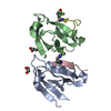
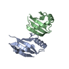
- Structure visualization
| Structure viewer | Molecule: MolmilJmol/JSmol |
|---|
- Downloads & links
-Download
| PDBx/mmCIF format | 4mjs.cif.gz | 414.5 KB | Display | PDBx/mmCIF format |
|---|---|---|---|---|
| PDB format | pdb4mjs.ent.gz | 341.5 KB | Display | PDB format |
| PDBx/mmJSON format | 4mjs.json.gz | Tree view | PDBx/mmJSON format | |
| Others |  Other downloads Other downloads |
-Validation report
| Arichive directory | https://data.pdbj.org/pub/pdb/validation_reports/mj/4mjsftp://data.pdbj.org/pub/pdb/validation_reports/mj/4mjs | HTTPS FTP |
|---|
-Related structure data
| Related structure data |  1wmhS S: Starting model for refinement |
|---|---|
| Similar structure data |
-Links
 PDBj
PDBj


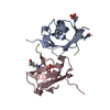
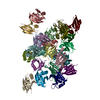
- Assembly


-Components
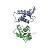
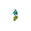
| #1: Protein | Mass: 10287.679 Da / Num. of mol.: 12 / Fragment: PB1 domain, UNP residues 15-101 Source method: isolated from a genetically manipulated source Source: (gene. exp.)  #2: Protein | Mass: 11542.182 Da / Num. of mol.: 12 / Fragment: PB1 domain, UNP residues 3-102 / Mutation: D69A,D71R Source method: isolated from a genetically manipulated source Source: (gene. exp.) Homo sapiens (human) / Gene: ORCA, OSIL, SQSTM1 / Plasmid: PACYCduet / Production host: #3: Chemical | ChemComp-EDO /   Mass: 62.068 Da / Num. of mol.: 4 / Source method: obtained synthetically / Formula: C2H6O2 Mass: 62.068 Da / Num. of mol.: 4 / Source method: obtained synthetically / Formula: C2H6O2#4: Water | ChemComp-HOH / |  Mass: 18.015 Da / Num. of mol.: 397 / Source method: isolated from a natural source / Formula: H2O Mass: 18.015 Da / Num. of mol.: 397 / Source method: isolated from a natural source / Formula: H2O |
|---|
-Experimental details
-Experiment
| Experiment | Method: X-RAY DIFFRACTION / Number of used crystals: 1 |
|---|
- Sample preparation
| Crystal | Density Matthews: 2.88 Å3/Da / Density % sol: 57.3 % |
|---|---|
| Crystal grow | Temperature: 294 K / Method: vapor diffusion, hanging drop / pH: 8.5 Details: 0.1M Tris-HCl, 8% PEG8000, 0.4M MgCl2, pH 8.5, VAPOR DIFFUSION, HANGING DROP, temperature 294K |
-Data collection
| Diffraction | Mean temperature: 100 K |
|---|---|
| Diffraction source | Source: SYNCHROTRON / Site: SSRF  / Beamline: BL17U / Wavelength: 0.97892 Å / Beamline: BL17U / Wavelength: 0.97892 Å |
| Detector | Type: ADSC QUANTUM 315r / Detector: CCD / Date: Dec 22, 2011 |
| Radiation | Monochromator: double crystal / Protocol: SINGLE WAVELENGTH / Monochromatic (M) / Laue (L): M / Scattering type: x-ray |
| Radiation wavelength | Wavelength: 0.97892 Å / Relative weight: 1 |
| Reflection | Resolution: 2.5→40 Å / Num. all: 105540 / Num. obs: 105401 / % possible obs: 100 % / Observed criterion σ(F): 5 / Observed criterion σ(I): 5 / Redundancy: 7.3 % / Rmerge(I) obs: 0.086 / Net I/σ(I): 22.6 |
| Reflection shell | Resolution: 2.5→2.59 Å / Redundancy: 7.5 % / Rmerge(I) obs: 0.519 / Mean I/σ(I) obs: 4.1 / % possible all: 100 |
- Processing
| Software |
| |||||||||||||||||||||||||||||||||||||||||||||||||||||||||||||||||||||||||||||||||||||||||||||||||||||||||||||||||||||||||||||||||||||||||||||||||||||||||||||||||||||||||||||||||||||||||||||||||||||||||||||||||||||||||
|---|---|---|---|---|---|---|---|---|---|---|---|---|---|---|---|---|---|---|---|---|---|---|---|---|---|---|---|---|---|---|---|---|---|---|---|---|---|---|---|---|---|---|---|---|---|---|---|---|---|---|---|---|---|---|---|---|---|---|---|---|---|---|---|---|---|---|---|---|---|---|---|---|---|---|---|---|---|---|---|---|---|---|---|---|---|---|---|---|---|---|---|---|---|---|---|---|---|---|---|---|---|---|---|---|---|---|---|---|---|---|---|---|---|---|---|---|---|---|---|---|---|---|---|---|---|---|---|---|---|---|---|---|---|---|---|---|---|---|---|---|---|---|---|---|---|---|---|---|---|---|---|---|---|---|---|---|---|---|---|---|---|---|---|---|---|---|---|---|---|---|---|---|---|---|---|---|---|---|---|---|---|---|---|---|---|---|---|---|---|---|---|---|---|---|---|---|---|---|---|---|---|---|---|---|---|---|---|---|---|---|---|---|---|---|---|---|---|---|
| Refinement | Method to determine structure: MOLECULAR REPLACEMENT Starting model: PDB ENTRY 1WMH Resolution: 2.5→39.534 Å / SU ML: 0.37 / σ(F): 1.34 / Phase error: 29.45 / Stereochemistry target values: ML
| |||||||||||||||||||||||||||||||||||||||||||||||||||||||||||||||||||||||||||||||||||||||||||||||||||||||||||||||||||||||||||||||||||||||||||||||||||||||||||||||||||||||||||||||||||||||||||||||||||||||||||||||||||||||||
| Solvent computation | Shrinkage radii: 0.98 Å / VDW probe radii: 1.2 Å / Solvent model: FLAT BULK SOLVENT MODEL / Bsol: 30.28 Å2 / ksol: 0.31 e/Å3 | |||||||||||||||||||||||||||||||||||||||||||||||||||||||||||||||||||||||||||||||||||||||||||||||||||||||||||||||||||||||||||||||||||||||||||||||||||||||||||||||||||||||||||||||||||||||||||||||||||||||||||||||||||||||||
| Displacement parameters |
| |||||||||||||||||||||||||||||||||||||||||||||||||||||||||||||||||||||||||||||||||||||||||||||||||||||||||||||||||||||||||||||||||||||||||||||||||||||||||||||||||||||||||||||||||||||||||||||||||||||||||||||||||||||||||
| Refinement step | Cycle: LAST / Resolution: 2.5→39.534 Å
| |||||||||||||||||||||||||||||||||||||||||||||||||||||||||||||||||||||||||||||||||||||||||||||||||||||||||||||||||||||||||||||||||||||||||||||||||||||||||||||||||||||||||||||||||||||||||||||||||||||||||||||||||||||||||
| Refine LS restraints |
| |||||||||||||||||||||||||||||||||||||||||||||||||||||||||||||||||||||||||||||||||||||||||||||||||||||||||||||||||||||||||||||||||||||||||||||||||||||||||||||||||||||||||||||||||||||||||||||||||||||||||||||||||||||||||
| LS refinement shell |
|
