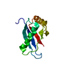
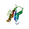
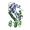
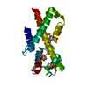
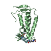
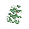
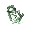
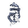
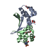
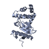
Entry Database : PDB / ID : 1wmhTitle Crystal structure of a PB1 domain complex of Protein kinase c iota and Par6 alpha Partitioning defective-6 homolog alpha Protein kinase C, iota type Keywords / / / / / / / Function / homology Function Domain/homology Component
/ / / / / / / / / / / / / / / / / / / / / / / / / / / / / / / / / / / / / / / / / / / / / / / / / / / / / / / / / / / / / / / / / / / / / / / / / / / / / / / / / / / / / / / / / / / / / / / / / / / / / / / / / / / / / / / / / / / / / / / / / / / / / / / / / / / / / / / / / / / / Biological species Homo sapiens (human)Method / / / Resolution : 1.5 Å Authors Hirano, Y. / Yoshinaga, S. / Suzuki, N.N. / Horiuchi, M. / Kohjima, M. / Takeya, R. / Sumimoto, H. / Inagaki, F. Journal : J.Biol.Chem. / Year : 2005Title : Structure of a Cell Polarity Regulator, a Complex between Atypical PKC and Par6 PB1 DomainsAuthors : Hirano, Y. / Yoshinaga, S. / Takeya, R. / Suzuki, N.N. / Horiuchi, M. / Kohjima, M. / Sumimoto, H. / Inagaki, F. History Deposition Jul 9, 2004 Deposition site / Processing site Revision 1.0 Dec 7, 2004 Provider / Type Revision 1.1 Apr 30, 2008 Group Revision 1.2 Jul 13, 2011 Group Revision 2.0 Mar 13, 2024 Group / Data collection / Database referencesCategory atom_site / chem_comp_atom ... atom_site / chem_comp_atom / chem_comp_bond / database_2 / struct_ref_seq_dif Item _atom_site.occupancy / _database_2.pdbx_DOI ... _atom_site.occupancy / _database_2.pdbx_DOI / _database_2.pdbx_database_accession / _struct_ref_seq_dif.details
Show all Show less
 Movie
Movie Controller

 Yorodumi
 Open data
 Basic information
 Components
 Keywords
 Function and homology information
 Homo sapiens (human)
 X-RAY DIFFRACTION /
  Authors
 Citation
 Structure visualization
 Downloads & links
 Other downloads
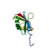


 PDBj
PDBj


 Assembly
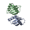

 Mass: 18.015 Da / Num. of mol.: 188 / Source method: isolated from a natural source / Formula: H2O
 Sample preparation
 / Beamline: BL41XU / Wavelength: 0.9791, 0.9793, 0.9795, 0.9843
 Processing