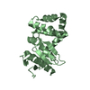
 Movie
Movie Controller


+ Open data

- Basic information
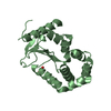
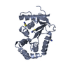
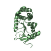
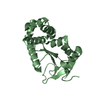
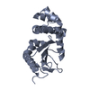
| Entry | Database: PDB / ID: 1a2j | ||||||
|---|---|---|---|---|---|---|---|
| Title | OXIDIZED DSBA CRYSTAL FORM II | ||||||
 Components Components | DISULFIDE BOND FORMATION PROTEIN | ||||||
 Keywords Keywords | OXIDOREDUCTASE / PROTEIN DISULFIDE ISOMERASE / PROTEIN FOLDING / REDOX PROTEIN / REDOX-ACTIVE CENTER | ||||||
| Function / homology |  Function and homology information Function and homology informationprotein disulfide isomerase activity / cellular response to antibiotic / Secretion of toxins / protein-disulfide reductase activity / outer membrane-bounded periplasmic space / periplasmic space / oxidoreductase activity Similarity search - Function | ||||||
| Biological species |  | ||||||
| Method |  X-RAY DIFFRACTION / MOLECULAR REPLACEMENT / Resolution: 2 Å X-RAY DIFFRACTION / MOLECULAR REPLACEMENT / Resolution: 2 Å | ||||||
 Authors Authors | Martin, J.L. / Guddat, L.W. | ||||||
 Citation Citation | Journal: Structure / Year: 1998 Title: Crystal structures of reduced and oxidized DsbA: investigation of domain motion and thiolate stabilization. Authors: Guddat, L.W. / Bardwell, J.C. / Martin, J.L. #1: Journal: Protein Sci. / Year: 1997Title: The Uncharged Surface Features Surrounding the Active Site of Escherichia Coli Dsba are Conserved and are Implicated in Peptide Binding Authors: Guddat, L.W. / Bardwell, J.C. / Zander, T. / Martin, J.L. #2: Journal: Protein Sci. / Year: 1997Title: Structural Analysis of Three His32 Mutants of Dsba: Support for an Electrostatic Role of His32 in Dsba Stability Authors: Guddat, L.W. / Bardwell, J.C. / Glockshuber, R. / Huber-Wunderlich, M. / Zander, T. / Martin, J.L. #3: Journal: Nature / Year: 1993Title: Crystal Structure of the Dsba Protein Required for Disulphide Bond Formation in Vivo Authors: Martin, J.L. / Bardwell, J.C. / Kuriyan, J. #4: Journal: J.Mol.Biol. / Year: 1993Title: Crystallization of Dsba, an Escherichia Coli Protein Required for Disulphide Bond Formation in Vivo Authors: Martin, J.L. / Waksman, G. / Bardwell, J.C. / Beckwith, J. / Kuriyan, J. | ||||||
| History |
|
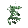
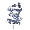
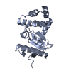
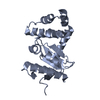
- Structure visualization
| Structure viewer | Molecule: MolmilJmol/JSmol |
|---|
- Downloads & links
-Download
| PDBx/mmCIF format | 1a2j.cif.gz | 50.3 KB | Display | PDBx/mmCIF format |
|---|---|---|---|---|
| PDB format | pdb1a2j.ent.gz | 35.8 KB | Display | PDB format |
| PDBx/mmJSON format | 1a2j.json.gz | Tree view | PDBx/mmJSON format | |
| Others |  Other downloads Other downloads |
-Validation report
| Arichive directory | https://data.pdbj.org/pub/pdb/validation_reports/a2/1a2jftp://data.pdbj.org/pub/pdb/validation_reports/a2/1a2j | HTTPS FTP |
|---|
-Related structure data
| Related structure data | 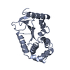 1a2lC 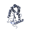 1a2mC  1fvkS S: Starting model for refinement C: citing same article ( |
|---|---|
| Similar structure data |


-Links
 PDBj
PDBj

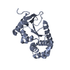
- Assembly
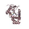
| Deposited unit | 
| ||||||||
|---|---|---|---|---|---|---|---|---|---|
| 1 |
| ||||||||
| Unit cell |
|
-Components
| #1: Protein | Mass: 21155.025 Da / Num. of mol.: 1 / Source method: isolated from a natural source / Source: (natural) |
|---|---|
| #2: Water | ChemComp-HOH /  Mass: 18.015 Da / Num. of mol.: 90 / Source method: isolated from a natural source / Formula: H2O Mass: 18.015 Da / Num. of mol.: 90 / Source method: isolated from a natural source / Formula: H2O |
| Has protein modification | Y |
-Experimental details
-Experiment
| Experiment | Method: X-RAY DIFFRACTION / Number of used crystals: 1 |
|---|
- Sample preparation
| Crystal | Density Matthews: 1.94 Å3/Da / Density % sol: 37 % | ||||||||||||||||||||
|---|---|---|---|---|---|---|---|---|---|---|---|---|---|---|---|---|---|---|---|---|---|
| Crystal grow | pH: 5 / Details: 27% PEG 4K IN 0.1M ACETATE BUFFER PH 5.0 | ||||||||||||||||||||
| Crystal grow | *PLUS Method: vapor diffusion, hanging drop | ||||||||||||||||||||
| Components of the solutions | *PLUS
|
-Data collection
| Diffraction | Mean temperature: 289 K |
|---|---|
| Diffraction source | Source: ROTATING ANODE / Type: RIGAKU RUH2R / Wavelength: 1.5418 |
| Detector | Type: RIGAKU / Detector: IMAGE PLATE / Date: Oct 10, 1995 / Details: YALE MIRRORS |
| Radiation | Monochromator: GRAPHITE(002) / Monochromatic (M) / Laue (L): M / Scattering type: x-ray |
| Radiation wavelength | Wavelength: 1.5418 Å / Relative weight: 1 |
| Reflection | Resolution: 2→50 Å / Num. obs: 38303 / % possible obs: 94.2 % / Observed criterion σ(I): 0 / Redundancy: 3.8 % / Biso Wilson estimate: 18.8 Å2 / Rmerge(I) obs: 0.046 / Rsym value: 0.046 / Net I/σ(I): 16 |
| Reflection shell | Resolution: 2→2.07 Å / Redundancy: 3.4 % / Rmerge(I) obs: 0.153 / Mean I/σ(I) obs: 7.4 / Rsym value: 0.153 / % possible all: 81.6 |
| Reflection | *PLUS Num. obs: 10434 / Num. measured all: 38303 |
| Reflection shell | *PLUS % possible obs: 81.6 % |
- Processing
| Software |
| ||||||||||||||||||||||||||||||||||||||||||||||||||||||||||||||||||||||||||||||||
|---|---|---|---|---|---|---|---|---|---|---|---|---|---|---|---|---|---|---|---|---|---|---|---|---|---|---|---|---|---|---|---|---|---|---|---|---|---|---|---|---|---|---|---|---|---|---|---|---|---|---|---|---|---|---|---|---|---|---|---|---|---|---|---|---|---|---|---|---|---|---|---|---|---|---|---|---|---|---|---|---|---|
| Refinement | Method to determine structure: MOLECULAR REPLACEMENT Starting model: PDB ENTRY 1FVK Resolution: 2→50 Å / Data cutoff high absF: 10000000 / Data cutoff low absF: 0.001 / σ(F): 1 Details: EIGHT RESIDUES GLU 4, GLU 13, LYS 48, LYS 98, GLU 120, LYS 132, GLN 146, AND LYS 183 WERE MODELLED AS ALANINE BECAUSE OF POORLY DEFINED SIDE CHAIN DENSITY. RESIDUES GLU 85, SER 106, SER 133, ...Details: EIGHT RESIDUES GLU 4, GLU 13, LYS 48, LYS 98, GLU 120, LYS 132, GLN 146, AND LYS 183 WERE MODELLED AS ALANINE BECAUSE OF POORLY DEFINED SIDE CHAIN DENSITY. RESIDUES GLU 85, SER 106, SER 133, AND SER 186 WERE MODELLED WITH TWO ALTERNATE CONFORMATIONS, EACH OF HALF OCCUPANCY. THE SIDE CHAINS OF GLU 13, GLU 85, SER 106, SER 133, AND SER 186 WERE MODELLED WITH TWO ALTERNATE CONFORMATIONS, EACH OF HALF OCCUPANCY.
| ||||||||||||||||||||||||||||||||||||||||||||||||||||||||||||||||||||||||||||||||
| Displacement parameters | Biso mean: 23.6 Å2 | ||||||||||||||||||||||||||||||||||||||||||||||||||||||||||||||||||||||||||||||||
| Refine analyze |
| ||||||||||||||||||||||||||||||||||||||||||||||||||||||||||||||||||||||||||||||||
| Refinement step | Cycle: LAST / Resolution: 2→50 Å
| ||||||||||||||||||||||||||||||||||||||||||||||||||||||||||||||||||||||||||||||||
| Refine LS restraints |
| ||||||||||||||||||||||||||||||||||||||||||||||||||||||||||||||||||||||||||||||||
| LS refinement shell | Resolution: 2→2.09 Å / Total num. of bins used: 8
| ||||||||||||||||||||||||||||||||||||||||||||||||||||||||||||||||||||||||||||||||
| Xplor file |
| ||||||||||||||||||||||||||||||||||||||||||||||||||||||||||||||||||||||||||||||||
| Software | *PLUS Name: X-PLOR / Version: 3.1 / Classification: refinement | ||||||||||||||||||||||||||||||||||||||||||||||||||||||||||||||||||||||||||||||||
| Refine LS restraints | *PLUS
|
