 Movie
Movie Controller

[English] 日本語
 Yorodumi
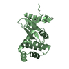
- PDB-4ocf: Crystal structure of the disulfide oxidoreductase DsbA (S30XXC33)... -
+ Open data

- Basic information
| Entry | Database: PDB / ID: 4ocf | ||||||
|---|---|---|---|---|---|---|---|
| Title | Crystal structure of the disulfide oxidoreductase DsbA (S30XXC33) active site mutant from Proteus mirabilis | ||||||
 Components Components | Thiol:disulfide interchange protein | ||||||
 Keywords Keywords | OXIDOREDUCTASE / Oxidative folding protein / virulence factor maturation protein / disulfide oxidoreductase / Thioredoxin / DsbA / Dithiol exchange / DsbB / Periplasmic | ||||||
| Function / homology |  Function and homology information Function and homology information | ||||||
| Biological species |  Proteus mirabilis (bacteria) Proteus mirabilis (bacteria) | ||||||
| Method |  X-RAY DIFFRACTION / SYNCHROTRON / MOLECULAR REPLACEMENT / Resolution: 1.979 Å X-RAY DIFFRACTION / SYNCHROTRON / MOLECULAR REPLACEMENT / Resolution: 1.979 Å | ||||||
 Authors Authors | Kurth, F. / Martin, J.L. | ||||||
 Citation Citation | Journal: J.Biol.Chem. / Year: 2014 Title: Crystal Structure of the Dithiol Oxidase DsbA Enzyme from Proteus Mirabilis Bound Non-covalently to an Active Site Peptide Ligand. Authors: Kurth, F. / Duprez, W. / Premkumar, L. / Schembri, M.A. / Fairlie, D.P. / Martin, J.L. | ||||||
| History |
|
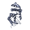
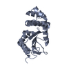
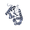
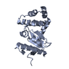
- Structure visualization
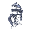
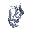
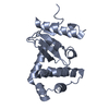
| Structure viewer | Molecule: MolmilJmol/JSmol |
|---|
- Downloads & links
-Download
| PDBx/mmCIF format | 4ocf.cif.gz | 292.9 KB | Display | PDBx/mmCIF format |
|---|---|---|---|---|
| PDB format | pdb4ocf.ent.gz | 241.8 KB | Display | PDB format |
| PDBx/mmJSON format | 4ocf.json.gz | Tree view | PDBx/mmJSON format | |
| Others |  Other downloads Other downloads |
-Validation report
| Arichive directory | https://data.pdbj.org/pub/pdb/validation_reports/oc/4ocfftp://data.pdbj.org/pub/pdb/validation_reports/oc/4ocf | HTTPS FTP |
|---|
-Related structure data
| Related structure data |  4oceSC  4od7C S: Starting model for refinement C: citing same article ( |
|---|---|
| Similar structure data |


-Links
 PDBj
PDBj


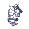
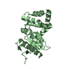
- Assembly
| Deposited unit | 
| ||||||||
|---|---|---|---|---|---|---|---|---|---|
| 1 | 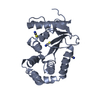
| ||||||||
| 2 | 
| ||||||||
| 3 | 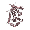
| ||||||||
| 4 | 
| ||||||||
| Unit cell |
|
-Components
| #1: Protein | Mass: 21011.627 Da / Num. of mol.: 4 / Mutation: C30S Source method: isolated from a genetically manipulated source Source: (gene. exp.) Proteus mirabilis (bacteria) / Strain: HI4320 / Gene: dsbA, PMI2828 / Production host: #2: Chemical | ChemComp-SCN /   Mass: 58.082 Da / Num. of mol.: 9 / Source method: obtained synthetically / Formula: CNS Mass: 58.082 Da / Num. of mol.: 9 / Source method: obtained synthetically / Formula: CNS#3: Water | ChemComp-HOH / |  Mass: 18.015 Da / Num. of mol.: 810 / Source method: isolated from a natural source / Formula: H2O Mass: 18.015 Da / Num. of mol.: 810 / Source method: isolated from a natural source / Formula: H2O |
|---|
-Experimental details
-Experiment
| Experiment | Method: X-RAY DIFFRACTION / Number of used crystals: 1 |
|---|
- Sample preparation
| Crystal | Density Matthews: 2.07 Å3/Da / Density % sol: 40.7 % |
|---|---|
| Crystal grow | Temperature: 293 K / Method: vapor diffusion, hanging drop Details: KSCN, PEG33500, VAPOR DIFFUSION, HANGING DROP, temperature 293K |
-Data collection
| Diffraction | Mean temperature: 100 K |
|---|---|
| Diffraction source | Source: SYNCHROTRON / Site: Australian Synchrotron  / Beamline: MX2 / Wavelength: 0.9537 Å / Beamline: MX2 / Wavelength: 0.9537 Å |
| Detector | Type: ADSC QUANTUM 315r / Detector: CCD / Date: Nov 8, 2012 |
| Radiation | Monochromator: si(111) / Protocol: SINGLE WAVELENGTH / Monochromatic (M) / Laue (L): M / Scattering type: x-ray |
| Radiation wavelength | Wavelength: 0.9537 Å / Relative weight: 1 |
| Reflection | Resolution: 1.979→57.22 Å / Num. all: 47865 / Num. obs: 47865 / % possible obs: 99.7 % / Observed criterion σ(F): 2 / Observed criterion σ(I): 2 / Redundancy: 3.7 % / Biso Wilson estimate: 12.34 Å2 / Rmerge(I) obs: 0.073 / Net I/σ(I): 14.3 |
| Reflection shell | Resolution: 1.979→2.09 Å / Redundancy: 3.6 % / Rmerge(I) obs: 0.155 / Mean I/σ(I) obs: 8.4 / % possible all: 98.9 |
- Processing
| Software |
| |||||||||||||||||||||||||||||||||||||||||||||||||||||||||||||||||||||||||||||||||||||||||||||||||||||||||
|---|---|---|---|---|---|---|---|---|---|---|---|---|---|---|---|---|---|---|---|---|---|---|---|---|---|---|---|---|---|---|---|---|---|---|---|---|---|---|---|---|---|---|---|---|---|---|---|---|---|---|---|---|---|---|---|---|---|---|---|---|---|---|---|---|---|---|---|---|---|---|---|---|---|---|---|---|---|---|---|---|---|---|---|---|---|---|---|---|---|---|---|---|---|---|---|---|---|---|---|---|---|---|---|---|---|---|
| Refinement | Method to determine structure: MOLECULAR REPLACEMENT Starting model: 4OCE Resolution: 1.979→57.215 Å / SU ML: 0.21 / σ(F): 1.36 / Phase error: 20.9 / Stereochemistry target values: ML
| |||||||||||||||||||||||||||||||||||||||||||||||||||||||||||||||||||||||||||||||||||||||||||||||||||||||||
| Solvent computation | Shrinkage radii: 0.9 Å / VDW probe radii: 1.11 Å / Solvent model: FLAT BULK SOLVENT MODEL | |||||||||||||||||||||||||||||||||||||||||||||||||||||||||||||||||||||||||||||||||||||||||||||||||||||||||
| Refinement step | Cycle: LAST / Resolution: 1.979→57.215 Å
| |||||||||||||||||||||||||||||||||||||||||||||||||||||||||||||||||||||||||||||||||||||||||||||||||||||||||
| Refine LS restraints |
| |||||||||||||||||||||||||||||||||||||||||||||||||||||||||||||||||||||||||||||||||||||||||||||||||||||||||
| LS refinement shell |
|
