 Movie
Movie Controller


+ Open data

- Basic information
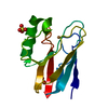
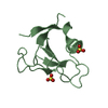
| Entry | Database: PDB / ID: 2fne | ||||||
|---|---|---|---|---|---|---|---|
| Title | The crystal structure of the 13th PDZ domain of MPDZ | ||||||
 Components Components | Multiple PDZ domain protein | ||||||
 Keywords Keywords | STRUCTURAL GENOMICS / UNKNOWN FUNCTION / Structural Protein / SGC / Structural Genomics Consortium | ||||||
| Function / homology |  Function and homology information Function and homology informationtight junction assembly / apicolateral plasma membrane / bicellular tight junction / apical part of cell / apical plasma membrane / postsynaptic density / dendrite / plasma membrane / cytoplasm Similarity search - Function | ||||||
| Biological species |  Homo sapiens (human) Homo sapiens (human) | ||||||
| Method |  X-RAY DIFFRACTION / SYNCHROTRON / MOLECULAR REPLACEMENT / Resolution: 1.83 Å X-RAY DIFFRACTION / SYNCHROTRON / MOLECULAR REPLACEMENT / Resolution: 1.83 Å | ||||||
 Authors Authors | Papagrigoriou, E. / Berridge, G. / Johansson, C. / Colebrook, S. / Salah, E. / Burgess, N. / Smee, C. / Savitsky, P. / Bray, J. / Schoch, G. ...Papagrigoriou, E. / Berridge, G. / Johansson, C. / Colebrook, S. / Salah, E. / Burgess, N. / Smee, C. / Savitsky, P. / Bray, J. / Schoch, G. / Phillips, C. / Gileadi, C. / Soundarajan, M. / Yang, X. / Elkins, J.M. / Gorrec, F. / Turnbull, A. / Edwards, A. / Arrowsmith, C. / Weigelt, J. / Sundstrom, M. / Doyle, D.A. / Structural Genomics Consortium (SGC) | ||||||
 Citation Citation | Journal: Protein Sci. / Year: 2007 Title: Structure of PICK1 and other PDZ domains obtained with the help of self-binding C-terminal extensions. Authors: Elkins, J.M. / Papagrigoriou, E. / Berridge, G. / Yang, X. / Phillips, C. / Gileadi, C. / Savitsky, P. / Doyle, D.A. | ||||||
| History |
|
- Structure visualization
| Structure viewer | Molecule: MolmilJmol/JSmol |
|---|
- Downloads & links
-Download
| PDBx/mmCIF format | 2fne.cif.gz | 73.3 KB | Display | PDBx/mmCIF format |
|---|---|---|---|---|
| PDB format | pdb2fne.ent.gz | 53.9 KB | Display | PDB format |
| PDBx/mmJSON format | 2fne.json.gz | Tree view | PDBx/mmJSON format | |
| Others |  Other downloads Other downloads |
-Validation report
| Arichive directory | https://data.pdbj.org/pub/pdb/validation_reports/fn/2fneftp://data.pdbj.org/pub/pdb/validation_reports/fn/2fne | HTTPS FTP |
|---|
-Related structure data
| Related structure data |  2bygSC  2fcfC 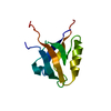 2gzvC  2he2C 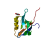 2he4C  2i1nC  2iwnC  2iwoC  2iwpC  2iwqC S: Starting model for refinement C: citing same article ( |
|---|---|
| Similar structure data |


-Links
 PDBj
PDBj- Assembly
| Deposited unit | 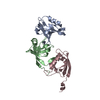
| ||||||||||||||||||||||||
|---|---|---|---|---|---|---|---|---|---|---|---|---|---|---|---|---|---|---|---|---|---|---|---|---|---|
| 1 | 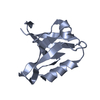
| ||||||||||||||||||||||||
| 2 | 
| ||||||||||||||||||||||||
| 3 | 
| ||||||||||||||||||||||||
| 4 | 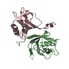
| ||||||||||||||||||||||||
| Unit cell |
| ||||||||||||||||||||||||
| Noncrystallographic symmetry (NCS) | NCS domain:
NCS domain segments: Component-ID: 1 / Ens-ID: 1 / Beg auth comp-ID: GLN / Beg label comp-ID: GLN / End auth comp-ID: VAL / End label comp-ID: VAL / Refine code: 4 / Auth seq-ID: 1956 - 2048 / Label seq-ID: 25 - 117
| ||||||||||||||||||||||||
| Details | Each of the three monomers found in the asu represents a biological unit |
-Components
| #1: Protein | Mass: 12529.081 Da / Num. of mol.: 3 / Fragment: MPDZ domain, residues 1955-2042 Source method: isolated from a genetically manipulated source Source: (gene. exp.) Homo sapiens (human) / Gene: MPDZ, MUPP1 / Plasmid: pNIC28-Bsa4 / Production host:  #2: Water | ChemComp-HOH / |  Mass: 18.015 Da / Num. of mol.: 199 / Source method: isolated from a natural source / Formula: H2O Mass: 18.015 Da / Num. of mol.: 199 / Source method: isolated from a natural source / Formula: H2O |
|---|
-Experimental details
-Experiment
| Experiment | Method: X-RAY DIFFRACTION / Number of used crystals: 1 |
|---|
- Sample preparation
| Crystal | Density Matthews: 2.1 Å3/Da / Density % sol: 40 % |
|---|---|
| Crystal grow | Temperature: 298 K / Method: vapor diffusion, sitting drop / pH: 4.6 Details: 8% PEG P3350, 0.1M acetate, pH 4.6, VAPOR DIFFUSION, SITTING DROP, temperature 298K |
-Data collection
| Diffraction | Mean temperature: 100 K |
|---|---|
| Diffraction source | Source: SYNCHROTRON / Site: SLS  / Beamline: X10SA / Wavelength: 0.979 Å / Beamline: X10SA / Wavelength: 0.979 Å |
| Detector | Type: MARRESEARCH / Detector: CCD / Date: Dec 9, 2005 |
| Radiation | Monochromator: Si(111) / Protocol: SINGLE WAVELENGTH / Monochromatic (M) / Laue (L): M / Scattering type: x-ray |
| Radiation wavelength | Wavelength: 0.979 Å / Relative weight: 1 |
| Reflection | Resolution: 1.83→39.5 Å / Num. all: 26633 / Num. obs: 26605 / % possible obs: 99.9 % / Observed criterion σ(F): 0 / Observed criterion σ(I): 0 / Rmerge(I) obs: 0.0508 |
| Reflection shell | Resolution: 1.83→1.95 Å / Rmerge(I) obs: 0.4753 / Mean I/σ(I) obs: 2.15 / Num. unique all: 4556 / % possible all: 100 |
- Processing
| Software |
| ||||||||||||||||||||||||||||||||||||||||||||||||||||||||||||||||||||||||||||||||||||||||||||||||||||||||||||||||||||||||||||||||||||||||||||||||||||||||||||||||||||||||||
|---|---|---|---|---|---|---|---|---|---|---|---|---|---|---|---|---|---|---|---|---|---|---|---|---|---|---|---|---|---|---|---|---|---|---|---|---|---|---|---|---|---|---|---|---|---|---|---|---|---|---|---|---|---|---|---|---|---|---|---|---|---|---|---|---|---|---|---|---|---|---|---|---|---|---|---|---|---|---|---|---|---|---|---|---|---|---|---|---|---|---|---|---|---|---|---|---|---|---|---|---|---|---|---|---|---|---|---|---|---|---|---|---|---|---|---|---|---|---|---|---|---|---|---|---|---|---|---|---|---|---|---|---|---|---|---|---|---|---|---|---|---|---|---|---|---|---|---|---|---|---|---|---|---|---|---|---|---|---|---|---|---|---|---|---|---|---|---|---|---|---|---|
| Refinement | Method to determine structure: MOLECULAR REPLACEMENT Starting model: 2byg Resolution: 1.83→39.5 Å / Cor.coef. Fo:Fc: 0.956 / Cor.coef. Fo:Fc free: 0.932 / SU B: 6.208 / SU ML: 0.099 / Cross valid method: THROUGHOUT / σ(F): 0 / ESU R: 0.142 / ESU R Free: 0.142 / Stereochemistry target values: MAXIMUM LIKELIHOOD / Details: HYDROGENS HAVE BEEN ADDED IN THE RIDING POSITIONS
| ||||||||||||||||||||||||||||||||||||||||||||||||||||||||||||||||||||||||||||||||||||||||||||||||||||||||||||||||||||||||||||||||||||||||||||||||||||||||||||||||||||||||||
| Solvent computation | Ion probe radii: 0.8 Å / Shrinkage radii: 0.8 Å / VDW probe radii: 1.4 Å / Solvent model: BABINET MODEL WITH MASK | ||||||||||||||||||||||||||||||||||||||||||||||||||||||||||||||||||||||||||||||||||||||||||||||||||||||||||||||||||||||||||||||||||||||||||||||||||||||||||||||||||||||||||
| Displacement parameters | Biso mean: 44.104 Å2
| ||||||||||||||||||||||||||||||||||||||||||||||||||||||||||||||||||||||||||||||||||||||||||||||||||||||||||||||||||||||||||||||||||||||||||||||||||||||||||||||||||||||||||
| Refinement step | Cycle: LAST / Resolution: 1.83→39.5 Å
| ||||||||||||||||||||||||||||||||||||||||||||||||||||||||||||||||||||||||||||||||||||||||||||||||||||||||||||||||||||||||||||||||||||||||||||||||||||||||||||||||||||||||||
| Refine LS restraints |
| ||||||||||||||||||||||||||||||||||||||||||||||||||||||||||||||||||||||||||||||||||||||||||||||||||||||||||||||||||||||||||||||||||||||||||||||||||||||||||||||||||||||||||
| Refine LS restraints NCS | Ens-ID: 1 / Number: 636 / Refine-ID: X-RAY DIFFRACTION
| ||||||||||||||||||||||||||||||||||||||||||||||||||||||||||||||||||||||||||||||||||||||||||||||||||||||||||||||||||||||||||||||||||||||||||||||||||||||||||||||||||||||||||
| LS refinement shell | Resolution: 1.83→1.87 Å / Total num. of bins used: 20
|
