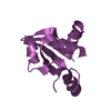
 Movie
Movie Controller


+ Open data

- Basic information
| Entry | Database: PDB / ID: 3nis | ||||||
|---|---|---|---|---|---|---|---|
| Title | The structure of UBR box (native2) | ||||||
 Components Components | E3 ubiquitin-protein ligase UBR1 | ||||||
 Keywords Keywords | METAL BINDING PROTEIN / E3 ubiquitin ligase / UBR box / Zinc-binding protein / N-end rule / Ligase | ||||||
| Function / homology |  Function and homology information Function and homology informationregulation of dipeptide transport / UBR1-RAD6 ubiquitin ligase complex / proteasome regulatory particle binding / stress-induced homeostatically regulated protein degradation pathway / ubiquitin-dependent protein catabolic process via the N-end rule pathway / mitochondria-associated ubiquitin-dependent protein catabolic process / cytoplasm protein quality control by the ubiquitin-proteasome system / proteasome regulatory particle, base subcomplex / ribosome-associated ubiquitin-dependent protein catabolic process / Antigen processing: Ubiquitination & Proteasome degradation ...regulation of dipeptide transport / UBR1-RAD6 ubiquitin ligase complex / proteasome regulatory particle binding / stress-induced homeostatically regulated protein degradation pathway / ubiquitin-dependent protein catabolic process via the N-end rule pathway / mitochondria-associated ubiquitin-dependent protein catabolic process / cytoplasm protein quality control by the ubiquitin-proteasome system / proteasome regulatory particle, base subcomplex / ribosome-associated ubiquitin-dependent protein catabolic process / Antigen processing: Ubiquitination & Proteasome degradation / cellular response to unfolded protein / protein monoubiquitination / ubiquitin ligase complex / ERAD pathway / RING-type E3 ubiquitin transferase / protein polyubiquitination / ubiquitin-protein transferase activity / ubiquitin protein ligase activity / protein ubiquitination / zinc ion binding / cytoplasm Similarity search - Function | ||||||
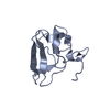
| Biological species |  | ||||||
| Method |  X-RAY DIFFRACTION / SYNCHROTRON / MOLECULAR REPLACEMENT / Resolution: 1.68 Å X-RAY DIFFRACTION / SYNCHROTRON / MOLECULAR REPLACEMENT / Resolution: 1.68 Å | ||||||
 Authors Authors | Choi, W.S. / Jeong, B.-C. / Lee, M.-R. / Song, H.K. | ||||||
 Citation Citation | Journal: Nat.Struct.Mol.Biol. / Year: 2010 Title: Structural basis for the recognition of N-end rule substrates by the UBR box of ubiquitin ligases Authors: Choi, W.S. / Jeong, B.-C. / Joo, Y.J. / Lee, M.-R. / Kim, J. / Eck, M.J. / Song, H.K. | ||||||
| History |
|
- Structure visualization
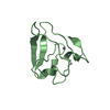
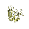
| Structure viewer | Molecule: MolmilJmol/JSmol |
|---|
- Downloads & links
-Download
| PDBx/mmCIF format | 3nis.cif.gz | 84.5 KB | Display | PDBx/mmCIF format |
|---|---|---|---|---|
| PDB format | pdb3nis.ent.gz | 62.9 KB | Display | PDB format |
| PDBx/mmJSON format | 3nis.json.gz | Tree view | PDBx/mmJSON format | |
| Others |  Other downloads Other downloads |
-Validation report
| Arichive directory | https://data.pdbj.org/pub/pdb/validation_reports/ni/3nisftp://data.pdbj.org/pub/pdb/validation_reports/ni/3nis | HTTPS FTP |
|---|
-Related structure data
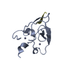
| Related structure data |  3nihC  3niiC  3nijC  3nikC  3nilC  3nimC  3ninC  3nitSC C: citing same article ( S: Starting model for refinement |
|---|---|
| Similar structure data |


-Links
 PDBj
PDBj


- Assembly
| Deposited unit | 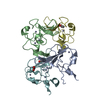
| ||||||||
|---|---|---|---|---|---|---|---|---|---|
| 1 | 
| ||||||||
| 2 | 
| ||||||||
| 3 | 
| ||||||||
| 4 | 
| ||||||||
| Unit cell |
|
-Components
| #1: Protein | Mass: 9236.394 Da / Num. of mol.: 4 / Fragment: UBR-type domain, residues 115-194 Source method: isolated from a genetically manipulated source Details: UBR box Source: (gene. exp.) Plasmid: pET / Production host:  #2: Chemical | ChemComp-ZN /   Mass: 65.409 Da / Num. of mol.: 12 / Source method: obtained synthetically / Formula: Zn Mass: 65.409 Da / Num. of mol.: 12 / Source method: obtained synthetically / Formula: Zn#3: Chemical |   Mass: 59.044 Da / Num. of mol.: 3 / Source method: obtained synthetically / Formula: C2H3O2 Mass: 59.044 Da / Num. of mol.: 3 / Source method: obtained synthetically / Formula: C2H3O2#4: Water | ChemComp-HOH / |  Mass: 18.015 Da / Num. of mol.: 262 / Source method: isolated from a natural source / Formula: H2O Mass: 18.015 Da / Num. of mol.: 262 / Source method: isolated from a natural source / Formula: H2O |
|---|
-Experimental details
-Experiment
| Experiment | Method: X-RAY DIFFRACTION / Number of used crystals: 1 |
|---|
- Sample preparation
| Crystal | Density Matthews: 2.17 Å3/Da / Density % sol: 43.43 % / Mosaicity: 0.475 ° |
|---|---|
| Crystal grow | Temperature: 295 K / Method: vapor diffusion, hanging drop / pH: 8 Details: 0.02M calcium chloride dehydrate, 0.1M sodium acetate trihydrate pH 4.8, 30%(v/v) MPD, VAPOR DIFFUSION, HANGING DROP, temperature 295K |
-Data collection
| Diffraction | Mean temperature: 100 K | ||||||||||||||||||||||||||||||||||||||||||||||||||||||||||||||||||
|---|---|---|---|---|---|---|---|---|---|---|---|---|---|---|---|---|---|---|---|---|---|---|---|---|---|---|---|---|---|---|---|---|---|---|---|---|---|---|---|---|---|---|---|---|---|---|---|---|---|---|---|---|---|---|---|---|---|---|---|---|---|---|---|---|---|---|---|
| Diffraction source | Source: SYNCHROTRON / Site: PAL/PLS  / Beamline: 6B / Wavelength: 1.1 Å / Beamline: 6B / Wavelength: 1.1 Å | ||||||||||||||||||||||||||||||||||||||||||||||||||||||||||||||||||
| Detector | Type: MAC Science DIP-2030 / Detector: IMAGE PLATE / Date: Feb 7, 2009 | ||||||||||||||||||||||||||||||||||||||||||||||||||||||||||||||||||
| Radiation | Protocol: SINGLE WAVELENGTH / Monochromatic (M) / Laue (L): M / Scattering type: x-ray | ||||||||||||||||||||||||||||||||||||||||||||||||||||||||||||||||||
| Radiation wavelength | Wavelength: 1.1 Å / Relative weight: 1 | ||||||||||||||||||||||||||||||||||||||||||||||||||||||||||||||||||
| Reflection | Resolution: 1.68→50 Å / Num. obs: 35555 / % possible obs: 100 % / Redundancy: 7.6 % / Rmerge(I) obs: 0.091 / Χ2: 1.793 / Net I/σ(I): 9.9 / Num. measured all: 270967 | ||||||||||||||||||||||||||||||||||||||||||||||||||||||||||||||||||
| Reflection shell |
|
- Processing
| Software |
| |||||||||||||||||||||||||||||||||||||||||||||||||||||||||||||||||||||||||||||||||||||||||||||||||||||||||||||||||||||||||||||
|---|---|---|---|---|---|---|---|---|---|---|---|---|---|---|---|---|---|---|---|---|---|---|---|---|---|---|---|---|---|---|---|---|---|---|---|---|---|---|---|---|---|---|---|---|---|---|---|---|---|---|---|---|---|---|---|---|---|---|---|---|---|---|---|---|---|---|---|---|---|---|---|---|---|---|---|---|---|---|---|---|---|---|---|---|---|---|---|---|---|---|---|---|---|---|---|---|---|---|---|---|---|---|---|---|---|---|---|---|---|---|---|---|---|---|---|---|---|---|---|---|---|---|---|---|---|---|
| Refinement | Method to determine structure: MOLECULAR REPLACEMENT Starting model: PDB ENTRY 3NIT Resolution: 1.68→25.94 Å / Cor.coef. Fo:Fc: 0.95 / Cor.coef. Fo:Fc free: 0.937 / Occupancy max: 1 / Occupancy min: 1 / SU B: 3.638 / SU ML: 0.081 / Cross valid method: THROUGHOUT / σ(F): 0 / ESU R Free: 0.11 / Stereochemistry target values: MAXIMUM LIKELIHOOD / Details: HYDROGENS HAVE BEEN ADDED IN THE RIDING POSITIONS
| |||||||||||||||||||||||||||||||||||||||||||||||||||||||||||||||||||||||||||||||||||||||||||||||||||||||||||||||||||||||||||||
| Solvent computation | Ion probe radii: 0.8 Å / Shrinkage radii: 0.8 Å / VDW probe radii: 1.2 Å / Solvent model: MASK | |||||||||||||||||||||||||||||||||||||||||||||||||||||||||||||||||||||||||||||||||||||||||||||||||||||||||||||||||||||||||||||
| Displacement parameters | Biso max: 103.03 Å2 / Biso mean: 25.003 Å2 / Biso min: 8.72 Å2
| |||||||||||||||||||||||||||||||||||||||||||||||||||||||||||||||||||||||||||||||||||||||||||||||||||||||||||||||||||||||||||||
| Refinement step | Cycle: LAST / Resolution: 1.68→25.94 Å
| |||||||||||||||||||||||||||||||||||||||||||||||||||||||||||||||||||||||||||||||||||||||||||||||||||||||||||||||||||||||||||||
| Refine LS restraints |
| |||||||||||||||||||||||||||||||||||||||||||||||||||||||||||||||||||||||||||||||||||||||||||||||||||||||||||||||||||||||||||||
| LS refinement shell | Resolution: 1.68→1.724 Å / Total num. of bins used: 20
| |||||||||||||||||||||||||||||||||||||||||||||||||||||||||||||||||||||||||||||||||||||||||||||||||||||||||||||||||||||||||||||
| Refinement TLS params. | Method: refined / Refine-ID: X-RAY DIFFRACTION
| |||||||||||||||||||||||||||||||||||||||||||||||||||||||||||||||||||||||||||||||||||||||||||||||||||||||||||||||||||||||||||||
| Refinement TLS group |
|
