 Movie
Movie Controller


+ Open data

- Basic information
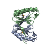
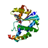
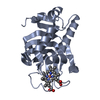
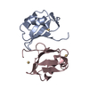
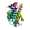
| Entry | Database: PDB / ID: 3zii | ||||||
|---|---|---|---|---|---|---|---|
| Title | Bacillus subtilis SepF G109K, C-terminal domain | ||||||
 Components Components | CELL DIVISION PROTEIN SEPF | ||||||
 Keywords Keywords | CELL CYCLE | ||||||
| Function / homology |  Function and homology information Function and homology informationcell septum assembly / division septum assembly / FtsZ-dependent cytokinesis / identical protein binding / cytoplasm Similarity search - Function | ||||||
| Biological species |  | ||||||
| Method |  X-RAY DIFFRACTION / SYNCHROTRON / MOLECULAR REPLACEMENT / Resolution: 2.2 Å X-RAY DIFFRACTION / SYNCHROTRON / MOLECULAR REPLACEMENT / Resolution: 2.2 Å | ||||||
 Authors Authors | Duman, R. / Ishikawa, S. / Celik, I. / Ogasawara, N. / Lowe, J. / Hamoen, L.W. | ||||||
 Citation Citation | Journal: Proc.Natl.Acad.Sci.USA / Year: 2013 Title: Structural and Genetic Analyses Reveal the Protein Sepf as a New Membrane Anchor for the Z Ring. Authors: Duman, R. / Ishikawa, S. / Celik, I. / Strahl, H. / Ogasawara, N. / Troc, P. / Lowe, J. / Hamoen, L.W. | ||||||
| History |
|
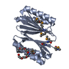
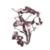
- Structure visualization
| Structure viewer | Molecule: MolmilJmol/JSmol |
|---|
- Downloads & links
-Download
| PDBx/mmCIF format | 3zii.cif.gz | 28.2 KB | Display | PDBx/mmCIF format |
|---|---|---|---|---|
| PDB format | pdb3zii.ent.gz | 17.9 KB | Display | PDB format |
| PDBx/mmJSON format | 3zii.json.gz | Tree view | PDBx/mmJSON format | |
| Others |  Other downloads Other downloads |
-Validation report
| Arichive directory | https://data.pdbj.org/pub/pdb/validation_reports/zi/3ziiftp://data.pdbj.org/pub/pdb/validation_reports/zi/3zii | HTTPS FTP |
|---|
-Related structure data


-Links
 PDBj
PDBj- Assembly
| Deposited unit | 
| ||||||||
|---|---|---|---|---|---|---|---|---|---|
| 1 | 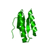
| ||||||||
| Unit cell |
|
-Components
| #1: Protein | Mass: 10747.072 Da / Num. of mol.: 1 / Fragment: C-TERMINAL DOMAIN, RESIDUES 57-151 / Mutation: YES Source method: isolated from a genetically manipulated source Source: (gene. exp.) |
|---|---|
| #2: Water | ChemComp-HOH /  Mass: 18.015 Da / Num. of mol.: 30 / Source method: isolated from a natural source / Formula: H2O Mass: 18.015 Da / Num. of mol.: 30 / Source method: isolated from a natural source / Formula: H2O |
-Experimental details
-Experiment
| Experiment | Method: X-RAY DIFFRACTION / Number of used crystals: 1 |
|---|
- Sample preparation
| Crystal | Density Matthews: 2.14 Å3/Da / Density % sol: 42.51 % / Description: NONE |
|---|---|
| Crystal grow | Details: 15% (V/V) ETHANOL, 0.1M TRIS PH 7.0 |
-Data collection
| Diffraction | Mean temperature: 100 K |
|---|---|
| Diffraction source | Source: SYNCHROTRON / Site: ESRF  / Beamline: ID23-1 / Wavelength: 0.9 / Beamline: ID23-1 / Wavelength: 0.9 |
| Detector | Date: Sep 4, 2011 |
| Radiation | Protocol: SINGLE WAVELENGTH / Monochromatic (M) / Laue (L): M / Scattering type: x-ray |
| Radiation wavelength | Wavelength: 0.9 Å / Relative weight: 1 |
| Reflection | Resolution: 2.2→51.82 Å / Num. obs: 4701 / % possible obs: 100 % / Observed criterion σ(I): 2 / Redundancy: 6.8 % / Biso Wilson estimate: 19.58 Å2 / Rmerge(I) obs: 0.15 / Net I/σ(I): 8.8 |
| Reflection shell | Resolution: 2.2→2.32 Å / Redundancy: 7 % / Rmerge(I) obs: 0.4 / Mean I/σ(I) obs: 4.5 / % possible all: 100 |
- Processing
| Software |
| ||||||||||||||||||||||||
|---|---|---|---|---|---|---|---|---|---|---|---|---|---|---|---|---|---|---|---|---|---|---|---|---|---|
| Refinement | Method to determine structure: MOLECULAR REPLACEMENT / Resolution: 2.2→25.91 Å / SU ML: 0.24 / σ(F): 1.4 / Phase error: 21.95 / Stereochemistry target values: ML
| ||||||||||||||||||||||||
| Solvent computation | Shrinkage radii: 0.86 Å / VDW probe radii: 1.1 Å / Solvent model: FLAT BULK SOLVENT MODEL / Bsol: 27.961 Å2 / ksol: 0.351 e/Å3 | ||||||||||||||||||||||||
| Displacement parameters |
| ||||||||||||||||||||||||
| Refinement step | Cycle: LAST / Resolution: 2.2→25.91 Å
| ||||||||||||||||||||||||
| Refine LS restraints |
| ||||||||||||||||||||||||
| LS refinement shell |
|
