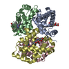
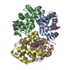
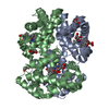
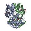
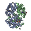
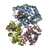
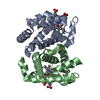
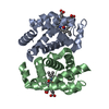
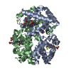
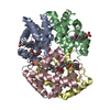
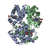
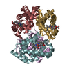
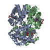
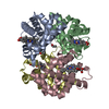
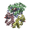
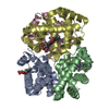
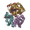
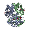
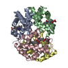
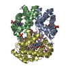
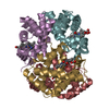
BIOMELECULE BIOLOGICAL ASSEMBLY IS A TETRAMER COMPOSED OF TWO ALPHA-BETA HETERODIMERS. HOWEVER, THE ...BIOMELECULE BIOLOGICAL ASSEMBLY IS A TETRAMER COMPOSED OF TWO ALPHA-BETA HETERODIMERS. HOWEVER, THE ASYMMETRIC UNIT CONTAINS TWO FULL HEMOGLOBIN TETRAMERS AND THEY REPRESENTS TWO DIFFERENT QUATERNARY STATES. ONE TETRAMER (CHAINS A,B,C,D) REPRESENTS THE NOVEL (R3) QUATERNARY STATE, ANOTHER TETRAMER (CHAINS E,F,G,H) IS IDENTICAL TO THE R2 QUATERNARY STATE (PDB ENTRY 1M9P). THE R3 QUATERNARY STATE IS IN PART STABILIZED BY PHOSPHATE ANION BOUND WITHIN THE CENTRAL CAVITY
Remark 999
SEQUENCE THE PROTEIN WAS NOT GENETICALLY MANIPULATED, BUT THE RESIDUE E6K OF CHAINS B,D,F AND H ARE ...SEQUENCE THE PROTEIN WAS NOT GENETICALLY MANIPULATED, BUT THE RESIDUE E6K OF CHAINS B,D,F AND H ARE ALLELIC VARIANTS OF HUMAN HEMOGLOBIN A.
BIOLOGICAL ASSEMBLY IS A TETRAMER COMPOSED OF TWO ALPHA-BETA HETERODIMERS. HOWEVER, THE ASYMMETRIC UNIT CONTAINS TWO FULL HEMOGLOBIN TETRAMERS AND THEY REPRESENTS TWO DIFFERENT QUATERNARY STATES. ONE TETRAMER (CHAINS A,B,C,D) REPRESENTS THE NOVEL (R3) QUATERNARY STATE, ANOTHER TETRAMER (CHAINS E,F,G,H) IS IDENTICAL TO THE R2 QUATERNARY STATE (PDB ENTRY 1M9P). THE R3 QUATERNARY STATE IS IN PART STABILIZED BY PHOSPHATE ANION BOUND WITHIN THE CENTRAL CAVITY
-
Components
-
Protein , 2 types, 8 molecules ACEGBDFH
#1: Protein
Hemoglobinalphachain
Mass: 15150.353 Da / Num. of mol.: 4 / Source method: isolated from a natural source / Source: (natural) Homo sapiens (human) / References: UniProt: P69905
#2: Protein
Hemoglobinbetachain
Mass: 15890.265 Da / Num. of mol.: 4 / Mutation: E6K / Source method: isolated from a natural source / Source: (natural) Homo sapiens (human) / References: UniProt: P68871
In the structure databanks used in Yorodumi, some data are registered as the other names, "COVID-19 virus" and "2019-nCoV". Here are the details of the virus and the list of structure data.
Jan 31, 2019. EMDB accession codes are about to change! (news from PDBe EMDB page)
EMDB accession codes are about to change! (news from PDBe EMDB page)
The allocation of 4 digits for EMDB accession codes will soon come to an end. Whilst these codes will remain in use, new EMDB accession codes will include an additional digit and will expand incrementally as the available range of codes is exhausted. The current 4-digit format prefixed with “EMD-” (i.e. EMD-XXXX) will advance to a 5-digit format (i.e. EMD-XXXXX), and so on. It is currently estimated that the 4-digit codes will be depleted around Spring 2019, at which point the 5-digit format will come into force.
The EM Navigator/Yorodumi systems omit the EMD- prefix.
Related info.:Q: What is EMD? / ID/Accession-code notation in Yorodumi/EM Navigator
Yorodumi is a browser for structure data from EMDB, PDB, SASBDB, etc.
This page is also the successor to EM Navigator detail page, and also detail information page/front-end page for Omokage search.
The word "yorodu" (or yorozu) is an old Japanese word meaning "ten thousand". "mi" (miru) is to see.
Related info.:EMDB / PDB / SASBDB / Comparison of 3 databanks / Yorodumi Search / Aug 31, 2016. New EM Navigator & Yorodumi / Yorodumi Papers / Jmol/JSmol / Function and homology information / Changes in new EM Navigator and Yorodumi
 Movie
Movie Controller

 Yorodumi
 Open data
 Basic information
 Components
 Keywords
 Function and homology information
 Homo sapiens (human)
 X-RAY DIFFRACTION /
  Authors
 Citation
 Structure visualization
 Downloads & links
 Other downloads
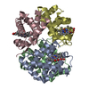


 PDBj
PDBj


 Assembly
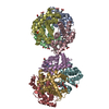


 Mass: 616.487 Da / Num. of mol.: 8 / Source method: obtained synthetically / Formula: C34H32FeN4O4
 Mass: 28.010 Da / Num. of mol.: 8 / Source method: obtained synthetically / Formula: CO
 Mass: 94.971 Da / Num. of mol.: 1 / Source method: obtained synthetically / Formula: PO4
 Sample preparation
 Processing