National Institutes of Health/National Heart, Lung, and Blood Institute (NIH/NHLBI)
P01-HL110900
United States
Robert A. Welch Foundation
C0612
United States
National Institute of Food and Agriculture (NIFA, United States)
United States
Citation
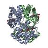
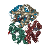
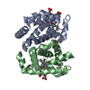
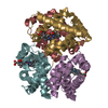
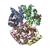
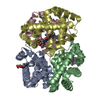
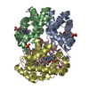
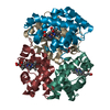
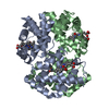
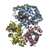
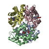
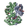
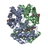
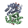
Journal: To Be Published Title: Structure of the Human Hemoglobin Mutant Hb Providence (A-Gly-C:V1M; B,D:V1M,K82D; Ferrous, carbonmonoxy bound) Authors: Soman, J. / Olson, J.S.
Mass: 18.015 Da / Num. of mol.: 164 / Source method: isolated from a natural source / Formula: H2O
-
Details
Sequence details
Gly142 was engineered into the sequence of the alpha chain to form a linker between the two alpha ...Gly142 was engineered into the sequence of the alpha chain to form a linker between the two alpha chains A and C.
-
Experimental details
-
Experiment
Experiment
Method: X-RAY DIFFRACTION / Number of used crystals: 1
-
Sample preparation
Crystal
Density Matthews: 2.19 Å3/Da / Density % sol: 43.82 %
In the structure databanks used in Yorodumi, some data are registered as the other names, "COVID-19 virus" and "2019-nCoV". Here are the details of the virus and the list of structure data.
Jan 31, 2019. EMDB accession codes are about to change! (news from PDBe EMDB page)
EMDB accession codes are about to change! (news from PDBe EMDB page)
The allocation of 4 digits for EMDB accession codes will soon come to an end. Whilst these codes will remain in use, new EMDB accession codes will include an additional digit and will expand incrementally as the available range of codes is exhausted. The current 4-digit format prefixed with “EMD-” (i.e. EMD-XXXX) will advance to a 5-digit format (i.e. EMD-XXXXX), and so on. It is currently estimated that the 4-digit codes will be depleted around Spring 2019, at which point the 5-digit format will come into force.
The EM Navigator/Yorodumi systems omit the EMD- prefix.
Related info.:Q: What is EMD? / ID/Accession-code notation in Yorodumi/EM Navigator
Yorodumi is a browser for structure data from EMDB, PDB, SASBDB, etc.
This page is also the successor to EM Navigator detail page, and also detail information page/front-end page for Omokage search.
The word "yorodu" (or yorozu) is an old Japanese word meaning "ten thousand". "mi" (miru) is to see.
Related info.:EMDB / PDB / SASBDB / Comparison of 3 databanks / Yorodumi Search / Aug 31, 2016. New EM Navigator & Yorodumi / Yorodumi Papers / Jmol/JSmol / Function and homology information / Changes in new EM Navigator and Yorodumi
 Movie
Movie Controller

 Yorodumi
 Open data
 Basic information
 Components
 Keywords
 Function and homology information
 Homo sapiens (human)
 X-RAY DIFFRACTION /
  Authors
 United States, 3items
  Citation
 Structure visualization
 Downloads & links
 Other downloads
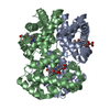


 PDBj
PDBj


 Assembly


 Mass: 616.487 Da / Num. of mol.: 2 / Source method: isolated from a natural source / Formula: C34H32FeN4O4
 Mass: 28.010 Da / Num. of mol.: 2 / Source method: obtained synthetically / Formula: CO
 Mass: 94.971 Da / Num. of mol.: 1 / Source method: obtained synthetically / Formula: PO4
 Sample preparation
 Processing