 Movie
Movie Controller


+ Open data

- Basic information
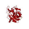
| Entry | Database: PDB / ID: 1rka | ||||||
|---|---|---|---|---|---|---|---|
| Title | THE APO FORM OF E. COLI RIBOKINASE | ||||||
 Components Components | PROTEIN (RIBOKINASE) | ||||||
 Keywords Keywords | TRANSFERASE / CARBOHYDRATE KINASE / RIBOSE / NUCLEOTIDE BINDING / INDUCED FIT | ||||||
| Function / homology |  Function and homology information Function and homology informationribokinase / ribokinase activity / D-ribose catabolic process / protein homodimerization activity / ATP binding / metal ion binding / cytosol Similarity search - Function | ||||||
| Biological species |  | ||||||
| Method |  X-RAY DIFFRACTION / SYNCHROTRON / MOLECULAR REPLACEMENT / Resolution: 2.3 Å X-RAY DIFFRACTION / SYNCHROTRON / MOLECULAR REPLACEMENT / Resolution: 2.3 Å | ||||||
 Authors Authors | Sigrell, J.A. / Cameron, A.D. / Mowbray, S.L. | ||||||
 Citation Citation | Journal: J.Mol.Biol. / Year: 1999 Title: Induced fit on sugar binding activates ribokinase. Authors: Sigrell, J.A. / Cameron, A.D. / Mowbray, S.L. #1: Journal: Structure / Year: 1998Title: Structure of Escherichia coli ribokinase in complex with ribose and dinucleotide determined to 1.8 A resolution: insights into a new family of kinase structures. Authors: Sigrell, J.A. / Cameron, A.D. / Jones, T.A. / Mowbray, S.L. #2: Journal: Protein Sci. / Year: 1997 Title: Purification, characterization, and crystallization of Escherichia coli ribokinase. Authors: Sigrell, J.A. / Cameron, A.D. / Jones, T.A. / Mowbray, S.L. #3: Journal: J.Biol.Chem. / Year: 1986 Title: Ribokinase from Escherichia coli K12. Nucleotide sequence and overexpression of the rbsK gene and purification of ribokinase. Authors: Hope, J.N. / Bell, A.W. / Hermodson, M.A. / Groarke, J.M. | ||||||
| History |
|
- Structure visualization
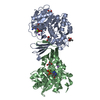
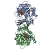
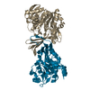
| Structure viewer | Molecule: MolmilJmol/JSmol |
|---|
- Downloads & links
-Download
| PDBx/mmCIF format | 1rka.cif.gz | 71.2 KB | Display | PDBx/mmCIF format |
|---|---|---|---|---|
| PDB format | pdb1rka.ent.gz | 52.4 KB | Display | PDB format |
| PDBx/mmJSON format | 1rka.json.gz | Tree view | PDBx/mmJSON format | |
| Others |  Other downloads Other downloads |
-Validation report
| Arichive directory | https://data.pdbj.org/pub/pdb/validation_reports/rk/1rkaftp://data.pdbj.org/pub/pdb/validation_reports/rk/1rka | HTTPS FTP |
|---|
-Related structure data
| Related structure data | 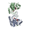 1rk2C 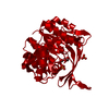 1rksC 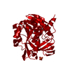 1rkdS S: Starting model for refinement C: citing same article ( |
|---|---|
| Similar structure data |


-Links
 PDBj
PDBj- Assembly
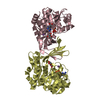
| Deposited unit | 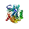
| ||||||||||
|---|---|---|---|---|---|---|---|---|---|---|---|
| 1 | 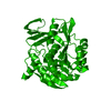
| ||||||||||
| Unit cell |
|
-Components
| #1: Protein | Mass: 32320.393 Da / Num. of mol.: 1 Source method: isolated from a genetically manipulated source Source: (gene. exp.) Description: THE RBSK GENE WAS CLONED BEHIND AURCE 11 TRP-PROMOTER, FORMING THE PLASMID PJGK10 Cellular location: CYTOPLASM / Gene: RBSK / Plasmid: PJGK10 / Production host: |
|---|---|
| #2: Water | ChemComp-HOH /  Mass: 18.015 Da / Num. of mol.: 123 / Source method: isolated from a natural source / Formula: H2O Mass: 18.015 Da / Num. of mol.: 123 / Source method: isolated from a natural source / Formula: H2O |
-Experimental details
-Experiment
| Experiment | Method: X-RAY DIFFRACTION / Number of used crystals: 1 |
|---|
- Sample preparation
| Crystal | Density Matthews: 2.1 Å3/Da / Density % sol: 40.2 % | ||||||||||||||||||||||||||||||||||||||||||
|---|---|---|---|---|---|---|---|---|---|---|---|---|---|---|---|---|---|---|---|---|---|---|---|---|---|---|---|---|---|---|---|---|---|---|---|---|---|---|---|---|---|---|---|
| Crystal grow | pH: 4.8 Details: CRYSTALS WERE GROWN IN 22-33% PEG 2000, MONO-METHYLETHER AS PRECIPITANT AND BUFFERED TO PH 4.8 WITH 0.1 M NA ACETATE. CRYO-PROTECTANT: MOTHER LIQUID ALSO CONTAINING 15% ETHYLENEGLYCOL. SOAK-TIME: 2-3 MINUTES. | ||||||||||||||||||||||||||||||||||||||||||
| Crystal grow | *PLUS Method: vapor diffusion, hanging dropDetails: drop consists of equal volume of protein and reservoir solutions | ||||||||||||||||||||||||||||||||||||||||||
| Components of the solutions | *PLUS
|
-Data collection
| Diffraction | Mean temperature: 100 K |
|---|---|
| Diffraction source | Source: SYNCHROTRON / Site: MAX II  / Beamline: I711 / Wavelength: 0.996 / Beamline: I711 / Wavelength: 0.996 |
| Detector | Type: MARRESEARCH / Detector: IMAGE PLATE / Date: May 15, 1998 / Details: MIRRORS |
| Radiation | Monochromator: SI(111) CRYSTAL / Protocol: SINGLE WAVELENGTH / Monochromatic (M) / Laue (L): M / Scattering type: x-ray |
| Radiation wavelength | Wavelength: 0.996 Å / Relative weight: 1 |
| Reflection | Resolution: 2.3→15 Å / Num. obs: 12644 / % possible obs: 96.4 % / Redundancy: 5.5 % / Biso Wilson estimate: 32 Å2 / Rmerge(I) obs: 0.056 / Net I/σ(I): 20.6 |
| Reflection shell | Resolution: 2.3→2.34 Å / Redundancy: 5.5 % / Rmerge(I) obs: 0.235 / Mean I/σ(I) obs: 6.7 / % possible all: 87.5 |
| Reflection | *PLUS Num. measured all: 62741 |
| Reflection shell | *PLUS % possible obs: 87.5 % |
- Processing
| Software |
| ||||||||||||||||||||||||||||||||||||||||||||||||||||||||||||||||||||||||||||||||
|---|---|---|---|---|---|---|---|---|---|---|---|---|---|---|---|---|---|---|---|---|---|---|---|---|---|---|---|---|---|---|---|---|---|---|---|---|---|---|---|---|---|---|---|---|---|---|---|---|---|---|---|---|---|---|---|---|---|---|---|---|---|---|---|---|---|---|---|---|---|---|---|---|---|---|---|---|---|---|---|---|---|
| Refinement | Method to determine structure: MOLECULAR REPLACEMENT Starting model: 1RKD: RESIDUES 4-9, 44-93, 123-241 AND 254-309. Resolution: 2.3→15 Å / Isotropic thermal model: RESTRAINED / Cross valid method: THROUGHOUT / σ(F): 0 / Stereochemistry target values: MLF USING AMPLITUDES
| ||||||||||||||||||||||||||||||||||||||||||||||||||||||||||||||||||||||||||||||||
| Solvent computation | Solvent model: EXPONENTIAL SCALING / Bsol: 45.9 Å2 / ksol: 0.33 e/Å3 | ||||||||||||||||||||||||||||||||||||||||||||||||||||||||||||||||||||||||||||||||
| Displacement parameters | Biso mean: 27 Å2
| ||||||||||||||||||||||||||||||||||||||||||||||||||||||||||||||||||||||||||||||||
| Refine analyze |
| ||||||||||||||||||||||||||||||||||||||||||||||||||||||||||||||||||||||||||||||||
| Refinement step | Cycle: LAST / Resolution: 2.3→15 Å
| ||||||||||||||||||||||||||||||||||||||||||||||||||||||||||||||||||||||||||||||||
| Refine LS restraints |
| ||||||||||||||||||||||||||||||||||||||||||||||||||||||||||||||||||||||||||||||||
| Xplor file |
| ||||||||||||||||||||||||||||||||||||||||||||||||||||||||||||||||||||||||||||||||
| Software | *PLUS Name: CNS / Version: 0.3 / Classification: refinement | ||||||||||||||||||||||||||||||||||||||||||||||||||||||||||||||||||||||||||||||||
| Refine LS restraints | *PLUS
|
