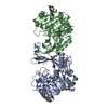
Entry Database : PDB / ID : 2c4eTitle Crystal Structure of Methanocaldococcus jannaschii Nucleoside Kinase - An Archaeal Member of the Ribokinase Family SUGAR KINASE MJ0406 Keywords / / / / Function / homology Function Domain/homology Component
/ / / / / / / / / / / / / / / / / / Biological species METHANOCOCCUS JANNASCHII (archaea)Method / / / Resolution : 1.7 Å Authors Arnfors, L. / Hansen, T. / Meining, W. / Schoenheit, P. / Ladenstein, R. Journal : Acta Crystallogr.,Sect.D / Year : 2006Title : Structure of Methanocaldococcus Jannaschii Nucleoside Kinase: An Archaeal Member of the Ribokinase Family.Authors : Arnfors, L. / Hansen, T. / Schoenheit, P. / Ladenstein, R. / Meining, W. History Deposition Oct 18, 2005 Deposition site / Processing site Revision 1.0 Aug 30, 2006 Provider / Type Revision 1.1 Jul 13, 2011 Group / Version format complianceRevision 1.2 Jan 17, 2018 Group / Category / Item Revision 1.3 May 8, 2024 Group Data collection / Database references ... Data collection / Database references / Derived calculations / Other Category chem_comp_atom / chem_comp_bond ... chem_comp_atom / chem_comp_bond / database_2 / pdbx_database_status / pdbx_struct_conn_angle / struct_conn / struct_site Item _database_2.pdbx_DOI / _database_2.pdbx_database_accession ... _database_2.pdbx_DOI / _database_2.pdbx_database_accession / _pdbx_database_status.status_code_sf / _pdbx_struct_conn_angle.ptnr1_auth_seq_id / _pdbx_struct_conn_angle.ptnr3_auth_seq_id / _pdbx_struct_conn_angle.value / _struct_conn.pdbx_dist_value / _struct_conn.ptnr2_auth_seq_id / _struct_site.pdbx_auth_asym_id / _struct_site.pdbx_auth_comp_id / _struct_site.pdbx_auth_seq_id
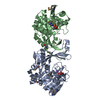
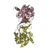
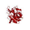
Show all Show less Remark 700 SHEET THE SHEET STRUCTURE OF THIS MOLECULE IS BIFURCATED. IN ORDER TO REPRESENT THIS FEATURE IN ... SHEET THE SHEET STRUCTURE OF THIS MOLECULE IS BIFURCATED. IN ORDER TO REPRESENT THIS FEATURE IN THE SHEET RECORDS BELOW, TWO SHEETS ARE DEFINED.
 Movie
Movie Controller

 Yorodumi
 Open data
 Basic information
 Components
 Keywords
 Function and homology information
 METHANOCOCCUS JANNASCHII (archaea)
 X-RAY DIFFRACTION /
  Authors
 Citation
 Structure visualization
 Downloads & links
 Other downloads
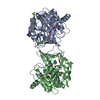


 PDBj
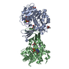
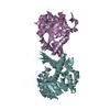
PDBj Assembly
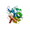


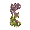
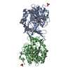
 Mass: 24.305 Da / Num. of mol.: 1 / Source method: obtained synthetically / Formula: Mg
 Mass: 18.015 Da / Num. of mol.: 113 / Source method: isolated from a natural source / Formula: H2O
 Sample preparation
 / Beamline: I711 / Wavelength: 1.134
  Processing