 Movie
Movie Controller


+ Open data

- Basic information
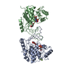
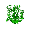
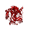
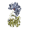
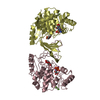
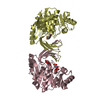
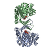
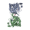
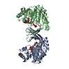
| Entry | Database: PDB / ID: 1rks | |||||||||
|---|---|---|---|---|---|---|---|---|---|---|
| Title | E. COLI RIBOKINASE IN COMPLEX WITH D-RIBOSE | |||||||||
 Components Components | PROTEIN (RIBOKINASE) | |||||||||
 Keywords Keywords | TRANSFERASE / CARBOHYDRATE KINASE / RIBOSE / INDUCED FIT | |||||||||
| Function / homology |  Function and homology information Function and homology informationribokinase / ribokinase activity / D-ribose catabolic process / protein homodimerization activity / ATP binding / metal ion binding / cytosol Similarity search - Function | |||||||||
| Biological species |  | |||||||||
| Method |  X-RAY DIFFRACTION / OTHER / Resolution: 2.4 Å X-RAY DIFFRACTION / OTHER / Resolution: 2.4 Å | |||||||||
 Authors Authors | Sigrell, J.A. / Cameron, A.D. / Mowbray, S.L. | |||||||||
 Citation Citation | Journal: J.Mol.Biol. / Year: 1999 Title: Induced fit on sugar binding activates ribokinase. Authors: Sigrell, J.A. / Cameron, A.D. / Mowbray, S.L. #1: Journal: Structure / Year: 1998Title: Structure of Escherichia Coli Ribokinase in Complex with Ribose and Nucleotide Determined to 1.8 A Resolution: Insights Into a New Family of Kinase Structures Authors: Sigrell, J.A. / Cameron, A.D. / Jones, T.A. / Mowbray, S.L. #2: Journal: Protein Sci. / Year: 1997Title: Purification, Characterization, and Crystallization of Escherichia Coli Ribokinase Authors: Sigrell, J.A. / Cameron, A.D. / Jones, T.A. / Mowbray, S.L. #3: Journal: J.Biol.Chem. / Year: 1986Title: Ribokinase from Escherichia Coli K12. Nucleotide Sequence and Overexpression of the Rbsk Gene and Purification of Ribokinase Authors: Hope, J.N. / Bell, A.W. / Hermodson, M.A. / Groarke, J.M. | |||||||||
| History |
|
- Structure visualization
| Structure viewer | Molecule: MolmilJmol/JSmol |
|---|
- Downloads & links
-Download
| PDBx/mmCIF format | 1rks.cif.gz | 69.9 KB | Display | PDBx/mmCIF format |
|---|---|---|---|---|
| PDB format | pdb1rks.ent.gz | 52.7 KB | Display | PDB format |
| PDBx/mmJSON format | 1rks.json.gz | Tree view | PDBx/mmJSON format | |
| Others |  Other downloads Other downloads |
-Validation report
| Arichive directory | https://data.pdbj.org/pub/pdb/validation_reports/rk/1rksftp://data.pdbj.org/pub/pdb/validation_reports/rk/1rks | HTTPS FTP |
|---|
-Related structure data


-Links
 PDBj
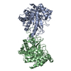
PDBj- Assembly
| Deposited unit | 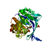
| ||||||||||
|---|---|---|---|---|---|---|---|---|---|---|---|
| 1 | 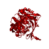
| ||||||||||
| Unit cell |
| ||||||||||
| Components on special symmetry positions |
|
-Components
| #1: Protein | Mass: 32320.393 Da / Num. of mol.: 1 Source method: isolated from a genetically manipulated source Source: (gene. exp.) Description: THE RBSK GENE WAS CLONED BEHIND AURCE 11 TRP-PROMOTER, FORMING THE PLASMID PJGK10 Cellular location: CYTOPLASM / Gene: RBSK / Plasmid: PJGK10 / Production host: | ||||
|---|---|---|---|---|---|
| #2: Sugar | ChemComp-RIB / 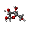  Type: D-saccharide, alpha linking / Mass: 150.130 Da / Num. of mol.: 1 Type: D-saccharide, alpha linking / Mass: 150.130 Da / Num. of mol.: 1Source method: isolated from a genetically manipulated source Formula: C5H10O5 | ||||
| #3: Chemical |   Mass: 94.971 Da / Num. of mol.: 2 / Source method: obtained synthetically / Formula: PO4 Mass: 94.971 Da / Num. of mol.: 2 / Source method: obtained synthetically / Formula: PO4#4: Water | ChemComp-HOH / |  Mass: 18.015 Da / Num. of mol.: 89 / Source method: isolated from a natural source / Formula: H2O Mass: 18.015 Da / Num. of mol.: 89 / Source method: isolated from a natural source / Formula: H2ONonpolymer details | THE SUGAR IS IN THE ALPHA-FURANOSE FORM AND HAS THE UNCOMMON O4'-ENDO PUCKERING. | |
-Experimental details
-Experiment
| Experiment | Method: X-RAY DIFFRACTION / Number of used crystals: 1 |
|---|
- Sample preparation
| Crystal | Density Matthews: 3.1 Å3/Da / Density % sol: 48.7 % / Description: RIGIDBODY REFINEMENT FROM 1RKD | ||||||||||||||||||||||||||||||||||||||||||||||||||||||||||||
|---|---|---|---|---|---|---|---|---|---|---|---|---|---|---|---|---|---|---|---|---|---|---|---|---|---|---|---|---|---|---|---|---|---|---|---|---|---|---|---|---|---|---|---|---|---|---|---|---|---|---|---|---|---|---|---|---|---|---|---|---|---|
| Crystal grow | Method: vapor diffusion, hanging drop / pH: 8.4 Details: 5 MM RIBOSE, 10 MM MGCL2 IN THE DROP, 0.1 M TRIS-HCL BUFFER, PH 8.4 WITH 2.1-2.4 M NH4H2PO4 AS PRECIPITANT, VAPOR DIFFUSION, HANGING DROP CRYO-SOLUTION: MOTHER LIQUOR CONTAINING 20% GLYCEROL. ...Details: 5 MM RIBOSE, 10 MM MGCL2 IN THE DROP, 0.1 M TRIS-HCL BUFFER, PH 8.4 WITH 2.1-2.4 M NH4H2PO4 AS PRECIPITANT, VAPOR DIFFUSION, HANGING DROP CRYO-SOLUTION: MOTHER LIQUOR CONTAINING 20% GLYCEROL. SOAK-TIME: 2-3 MINUTES. | ||||||||||||||||||||||||||||||||||||||||||||||||||||||||||||
| Crystal | *PLUS | ||||||||||||||||||||||||||||||||||||||||||||||||||||||||||||
| Crystal grow | *PLUS Temperature: 20 ℃ | ||||||||||||||||||||||||||||||||||||||||||||||||||||||||||||
| Components of the solutions | *PLUS
|
-Data collection
| Diffraction | Mean temperature: 100 K |
|---|---|
| Diffraction source | Source: ROTATING ANODE / Type: RIGAKU RU200 / Wavelength: 1.5418 |
| Detector | Type: RIGAKU RAXIS II / Detector: IMAGE PLATE / Date: Nov 15, 1997 / Details: MSC MIRRORS |
| Radiation | Monochromator: NI FILTER / Protocol: SINGLE WAVELENGTH / Monochromatic (M) / Laue (L): M / Scattering type: x-ray |
| Radiation wavelength | Wavelength: 1.5418 Å / Relative weight: 1 |
| Reflection | Resolution: 2.4→29 Å / Num. obs: 16814 / % possible obs: 99.9 % / Redundancy: 10.5 % / Biso Wilson estimate: 40 Å2 / Rmerge(I) obs: 0.069 / Net I/σ(I): 30.2 |
| Reflection shell | Resolution: 2.4→2.44 Å / Redundancy: 7.5 % / Rmerge(I) obs: 0.354 / Mean I/σ(I) obs: 5.8 / % possible all: 99.8 |
| Reflection | *PLUS Num. measured all: 134333 |
| Reflection shell | *PLUS % possible obs: 99.8 % |
- Processing
| Software |
| ||||||||||||||||||||||||||||||||||||||||||||||||||||||||||||||||||||||||||||||||||||
|---|---|---|---|---|---|---|---|---|---|---|---|---|---|---|---|---|---|---|---|---|---|---|---|---|---|---|---|---|---|---|---|---|---|---|---|---|---|---|---|---|---|---|---|---|---|---|---|---|---|---|---|---|---|---|---|---|---|---|---|---|---|---|---|---|---|---|---|---|---|---|---|---|---|---|---|---|---|---|---|---|---|---|---|---|---|
| Refinement | Method to determine structure: OTHER / Resolution: 2.4→15 Å / SU B: 7.72 / SU ML: 0.19 / Cross valid method: THROUGHOUT / σ(F): 0 / ESU R: 0.37 / ESU R Free: 0.28 / Details: BULK SOLVENT CORRECTION APPLIED
| ||||||||||||||||||||||||||||||||||||||||||||||||||||||||||||||||||||||||||||||||||||
| Displacement parameters | Biso mean: 45.3 Å2 | ||||||||||||||||||||||||||||||||||||||||||||||||||||||||||||||||||||||||||||||||||||
| Refinement step | Cycle: LAST / Resolution: 2.4→15 Å
| ||||||||||||||||||||||||||||||||||||||||||||||||||||||||||||||||||||||||||||||||||||
| Refine LS restraints |
| ||||||||||||||||||||||||||||||||||||||||||||||||||||||||||||||||||||||||||||||||||||
| Software | *PLUS Name: REFMAC / Classification: refinement | ||||||||||||||||||||||||||||||||||||||||||||||||||||||||||||||||||||||||||||||||||||
| Refinement | *PLUS Highest resolution: 2.4 Å / σ(F): 0 / % reflection Rfree: 11.1 % / Rfactor all: 0.231 / Rfactor obs: 0.229 | ||||||||||||||||||||||||||||||||||||||||||||||||||||||||||||||||||||||||||||||||||||
| Solvent computation | *PLUS | ||||||||||||||||||||||||||||||||||||||||||||||||||||||||||||||||||||||||||||||||||||
| Displacement parameters | *PLUS Biso mean: 45.3 Å2 | ||||||||||||||||||||||||||||||||||||||||||||||||||||||||||||||||||||||||||||||||||||
| Refine LS restraints | *PLUS
|
