 Movie
Movie Controller


+ Open data

- Basic information
| Entry | Database: PDB / ID: 6znx | ||||||
|---|---|---|---|---|---|---|---|
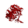
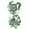
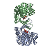
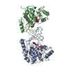
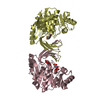
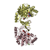
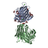
| Title | Ribokinase from Thermus Species | ||||||
 Components Components | Ribokinase | ||||||
 Keywords Keywords | TRANSFERASE / ADP | ||||||
| Function / homology |  Function and homology information Function and homology informationribokinase / ribokinase activity / D-ribose catabolic process / ATP binding / metal ion binding / cytosol Similarity search - Function | ||||||
| Biological species |   Thermus sp. 2.9 (bacteria) Thermus sp. 2.9 (bacteria) | ||||||
| Method |  X-RAY DIFFRACTION / SYNCHROTRON / MOLECULAR REPLACEMENT / Resolution: 2.4 Å X-RAY DIFFRACTION / SYNCHROTRON / MOLECULAR REPLACEMENT / Resolution: 2.4 Å | ||||||
 Authors Authors | Timofeev, V.I. / Abramchik, Y.A. / Tuzova, E.S. / Esipova, L.V. / Mikheeva, O.O. / Kostromina, M.A. / Kuranova, I.P. / Esipov, R.S. | ||||||
 Citation Citation | Journal: To Be Published Title: Ribokinase from Thermus Species Authors: Timofeev, V.I. / Abramchik, Y.A. / Tuzova, E.S. / Esipova, L.V. / Mikheeva, O.O. / Kostromina, M.A. / Kuranova, I.P. / Esipov, R.S. | ||||||
| History |
|
- Structure visualization
| Structure viewer | Molecule: MolmilJmol/JSmol |
|---|
- Downloads & links
-Download
| PDBx/mmCIF format | 6znx.cif.gz | 220.5 KB | Display | PDBx/mmCIF format |
|---|---|---|---|---|
| PDB format | pdb6znx.ent.gz | 177.1 KB | Display | PDB format |
| PDBx/mmJSON format | 6znx.json.gz | Tree view | PDBx/mmJSON format | |
| Others |  Other downloads Other downloads |
-Validation report
| Arichive directory | https://data.pdbj.org/pub/pdb/validation_reports/zn/6znxftp://data.pdbj.org/pub/pdb/validation_reports/zn/6znx | HTTPS FTP |
|---|
-Related structure data
| Related structure data | 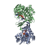 4xdaS S: Starting model for refinement |
|---|---|
| Similar structure data |


-Links
 PDBj
PDBj- Assembly
| Deposited unit | 
| ||||||||
|---|---|---|---|---|---|---|---|---|---|
| 1 | 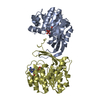
| ||||||||
| 2 | 
| ||||||||
| Unit cell |
|
-Components
| #1: Protein | Mass: 31026.439 Da / Num. of mol.: 4 Source method: isolated from a genetically manipulated source Source: (gene. exp.) Thermus sp. 2.9 (bacteria) / Gene: rbsK, QT17_05185 / Production host: #2: Chemical | ChemComp-ADP /   Mass: 427.201 Da / Num. of mol.: 4 / Source method: obtained synthetically / Formula: C10H15N5O10P2 / Comment: ADP, energy-carrying molecule*YM Mass: 427.201 Da / Num. of mol.: 4 / Source method: obtained synthetically / Formula: C10H15N5O10P2 / Comment: ADP, energy-carrying molecule*YM#3: Water | ChemComp-HOH / |  Mass: 18.015 Da / Num. of mol.: 186 / Source method: isolated from a natural source / Formula: H2O Mass: 18.015 Da / Num. of mol.: 186 / Source method: isolated from a natural source / Formula: H2OHas ligand of interest | N | |
|---|
-Experimental details
-Experiment
| Experiment | Method: X-RAY DIFFRACTION / Number of used crystals: 1 |
|---|
- Sample preparation
| Crystal | Density Matthews: 2.28 Å3/Da / Density % sol: 45.99 % |
|---|---|
| Crystal grow | Temperature: 293 K / Method: counter-diffusion / Details: PEG |
-Data collection
| Diffraction | Mean temperature: 100 K / Serial crystal experiment: N |
|---|---|
| Diffraction source | Source: SYNCHROTRON / Site: SPring-8  / Beamline: BL41XU / Wavelength: 0.8 Å / Beamline: BL41XU / Wavelength: 0.8 Å |
| Detector | Type: DECTRIS EIGER X 16M / Detector: PIXEL / Date: Oct 10, 2019 |
| Radiation | Protocol: SINGLE WAVELENGTH / Monochromatic (M) / Laue (L): M / Scattering type: x-ray |
| Radiation wavelength | Wavelength: 0.8 Å / Relative weight: 1 |
| Reflection | Resolution: 2.4→20 Å / Num. obs: 41928 / % possible obs: 96.91 % / Redundancy: 3.18 % / Rmerge(I) obs: 0.077 / Net I/σ(I): 5.2958 |
| Reflection shell | Resolution: 2.4→2.53 Å / Redundancy: 3.28 % / Rmerge(I) obs: 0.14 / Mean I/σ(I) obs: 4.47 / Num. unique obs: 6150 / % possible all: 97.73 |
- Processing
| Software |
| ||||||||||||||||||||||||||||||||||||||||||||||||||||||||||||
|---|---|---|---|---|---|---|---|---|---|---|---|---|---|---|---|---|---|---|---|---|---|---|---|---|---|---|---|---|---|---|---|---|---|---|---|---|---|---|---|---|---|---|---|---|---|---|---|---|---|---|---|---|---|---|---|---|---|---|---|---|---|
| Refinement | Method to determine structure: MOLECULAR REPLACEMENT Starting model: 4XDA Resolution: 2.4→20 Å / Cor.coef. Fo:Fc: 0.915 / Cor.coef. Fo:Fc free: 0.882 / SU B: 9.44 / SU ML: 0.224 / Cross valid method: THROUGHOUT / σ(F): 0 / ESU R: 0.697 / ESU R Free: 0.31 / Stereochemistry target values: MAXIMUM LIKELIHOOD Details: HYDROGENS HAVE BEEN ADDED IN THE RIDING POSITIONS U VALUES : REFINED INDIVIDUALLY
| ||||||||||||||||||||||||||||||||||||||||||||||||||||||||||||
| Solvent computation | Ion probe radii: 0.8 Å / Shrinkage radii: 0.8 Å / VDW probe radii: 1.2 Å / Solvent model: MASK | ||||||||||||||||||||||||||||||||||||||||||||||||||||||||||||
| Displacement parameters | Biso max: 107.21 Å2 / Biso mean: 36.213 Å2 / Biso min: 3.4 Å2
| ||||||||||||||||||||||||||||||||||||||||||||||||||||||||||||
| Refinement step | Cycle: final / Resolution: 2.4→20 Å
| ||||||||||||||||||||||||||||||||||||||||||||||||||||||||||||
| Refine LS restraints |
| ||||||||||||||||||||||||||||||||||||||||||||||||||||||||||||
| LS refinement shell | Resolution: 2.4→2.461 Å / Rfactor Rfree error: 0 / Total num. of bins used: 20
|
