 Movie
Movie Controller


+ Open data

- Basic information
| Entry | Database: PDB / ID: 1gqt | ||||||||||||
|---|---|---|---|---|---|---|---|---|---|---|---|---|---|
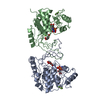
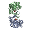
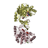
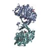
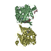
| Title | Activation of Ribokinase by Monovalent Cations | ||||||||||||
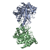
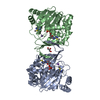
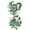
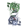
 Components Components | RIBOKINASE | ||||||||||||
 Keywords Keywords | TRANSFERASE / CARBOHYDRATE KINASE / RIBOSE / INDUCED FIT / BINDING OF MONOVALENT CATIONS / ACTIVATION BY MONOVALENT CATIONS | ||||||||||||
| Function / homology |  Function and homology information Function and homology informationribokinase / ribokinase activity / D-ribose catabolic process / protein homodimerization activity / ATP binding / metal ion binding / cytosol Similarity search - Function | ||||||||||||
| Biological species |  | ||||||||||||
| Method |  X-RAY DIFFRACTION / SYNCHROTRON / OTHER / Resolution: 2.34 Å X-RAY DIFFRACTION / SYNCHROTRON / OTHER / Resolution: 2.34 Å | ||||||||||||
 Authors Authors | Andersson, C.E. / Mowbray, S.L. | ||||||||||||
 Citation Citation | Journal: J.Mol.Biol. / Year: 2002 Title: Activation of Ribokinase by Monovalent Cations. Authors: Andersson, C.E. / Mowbray, S.L. #1: Journal: Structure / Year: 1998Title: Structure of Escherichia Coli Ribokinase in Complex with Ribose and Dinucleotide Determined to 1.8 A Resolution: Insights Into a New Family of Kinase Structures. Authors: Sigrell, J.A. / Cameron, A.D. / Jones, T.A. / Mowbray, S.L. #2: Journal: J.Biol.Chem. / Year: 1986 Title: Ribokinase from Escherichia Coli K12. Nucleotide Sequence and Overexpression of the Rbsk Gene and Purification of Ribokinase Authors: Hope, J.N. / Bell, A.W. / Hermodson, M.A. / Groarke, J.M. #3: Journal: J. Mol. Biol. / Year: 1999Title: Induced fit on sugar binding activates ribokinase. Authors: Sigrell, J.A. / Cameron, A.D. / Mowbray, S.L. | ||||||||||||
| History |
| ||||||||||||
| Remark 700 | SHEET THE SHEET STRUCTURE OF THIS MOLECULE IS BIFURCATED. IN ORDER TO REPRESENT THIS FEATURE IN ... SHEET THE SHEET STRUCTURE OF THIS MOLECULE IS BIFURCATED. IN ORDER TO REPRESENT THIS FEATURE IN THE SHEET RECORDS BELOW, TWO SHEETS ARE DEFINED. |
- Structure visualization
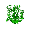
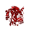
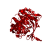
| Structure viewer | Molecule: MolmilJmol/JSmol |
|---|
- Downloads & links
-Download
| PDBx/mmCIF format | 1gqt.cif.gz | 241.4 KB | Display | PDBx/mmCIF format |
|---|---|---|---|---|
| PDB format | pdb1gqt.ent.gz | 193.6 KB | Display | PDB format |
| PDBx/mmJSON format | 1gqt.json.gz | Tree view | PDBx/mmJSON format | |
| Others |  Other downloads Other downloads |
-Validation report
| Arichive directory | https://data.pdbj.org/pub/pdb/validation_reports/gq/1gqtftp://data.pdbj.org/pub/pdb/validation_reports/gq/1gqt | HTTPS FTP |
|---|
-Related structure data
| Related structure data | |
|---|---|
| Similar structure data |


-Links
 PDBj
PDBj


- Assembly
| Deposited unit | 
| ||||||||
|---|---|---|---|---|---|---|---|---|---|
| 1 | 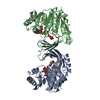
| ||||||||
| 2 | 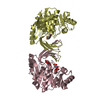
| ||||||||
| Unit cell |
|
-Components
| #1: Protein | Mass: 32320.393 Da / Num. of mol.: 4 Source method: isolated from a genetically manipulated source Source: (gene. exp.) References: UniProt: P05054, UniProt: P0A9J6*PLUS, ribokinase #2: Chemical | ChemComp-CS /   Mass: 132.905 Da / Num. of mol.: 4 / Source method: obtained synthetically / Formula: Cs Mass: 132.905 Da / Num. of mol.: 4 / Source method: obtained synthetically / Formula: Cs#3: Chemical |   Mass: 505.208 Da / Num. of mol.: 3 / Source method: obtained synthetically / Formula: C11H18N5O12P3 / Comment: AMP-PCP, energy-carrying molecule analogue*YM Mass: 505.208 Da / Num. of mol.: 3 / Source method: obtained synthetically / Formula: C11H18N5O12P3 / Comment: AMP-PCP, energy-carrying molecule analogue*YM#4: Sugar | ChemComp-RIB / 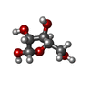  Type: D-saccharide, alpha linking / Mass: 150.130 Da / Num. of mol.: 4 Type: D-saccharide, alpha linking / Mass: 150.130 Da / Num. of mol.: 4Source method: isolated from a genetically manipulated source Formula: C5H10O5 #5: Water | ChemComp-HOH / |  Mass: 18.015 Da / Num. of mol.: 242 / Source method: isolated from a natural source / Formula: H2O Mass: 18.015 Da / Num. of mol.: 242 / Source method: isolated from a natural source / Formula: H2OCompound details | COMPOUND | |
|---|
-Experimental details
-Experiment
| Experiment | Method: X-RAY DIFFRACTION / Number of used crystals: 1 |
|---|
- Sample preparation
| Crystal | Density Matthews: 2.2 Å3/Da / Density % sol: 44.6 % Description: COORDINATES FROM 1RK2 WERE USED DIRECTLY IN REFINMENT. S.A. IN CNS WAS USED TO REMOVE MODEL BIAS. | |||||||||||||||||||||||||||||||||||||||||||||||||||||||||||||||
|---|---|---|---|---|---|---|---|---|---|---|---|---|---|---|---|---|---|---|---|---|---|---|---|---|---|---|---|---|---|---|---|---|---|---|---|---|---|---|---|---|---|---|---|---|---|---|---|---|---|---|---|---|---|---|---|---|---|---|---|---|---|---|---|---|
| Crystal grow | pH: 4.8 Details: CRYSTALS WERE GROWN IN DROPS CONTAINING 0.3 MM WILD-TYPE RK, 5 MM D-RIBOSE, 10 MM AMP-PCP, 75 MM MGCL2, 60 MM CSCL, 10% 2-METHYL-2,4-PENTANEDIOL, 10% POLYETHYLENE GLYCOL 4000 AND 50 MM SODIUM ACETATE, PH 4.8. | |||||||||||||||||||||||||||||||||||||||||||||||||||||||||||||||
| Crystal grow | *PLUS Temperature: 4 ℃ / Method: vapor diffusion, hanging drop / pH: 4.8 | |||||||||||||||||||||||||||||||||||||||||||||||||||||||||||||||
| Components of the solutions | *PLUS
|
-Data collection
| Diffraction | Mean temperature: 100 K |
|---|---|
| Diffraction source | Source: SYNCHROTRON / Site: ESRF  / Beamline: ID14-1 / Wavelength: 0.934 / Beamline: ID14-1 / Wavelength: 0.934 |
| Detector | Type: MARRESEARCH / Detector: CCD / Date: Feb 26, 2000 |
| Radiation | Protocol: SINGLE WAVELENGTH / Monochromatic (M) / Laue (L): M / Scattering type: x-ray |
| Radiation wavelength | Wavelength: 0.934 Å / Relative weight: 1 |
| Reflection | Resolution: 2.34→17 Å / Num. obs: 50296 / % possible obs: 98.2 % / Redundancy: 4.9 % / Rmerge(I) obs: 0.091 / Net I/σ(I): 10.9 |
| Reflection shell | Resolution: 2.34→2.38 Å / Rmerge(I) obs: 0.377 / Mean I/σ(I) obs: 3.8 / % possible all: 99.9 |
| Reflection | *PLUS Num. measured all: 245133 |
| Reflection shell | *PLUS % possible obs: 99.9 % |
- Processing
| Software |
| ||||||||||||||||||||||||||||||||||||||||||||||||||||||||||||||||||||||||||||||||||||||||||||||||||||||||||||||||||||||||||||||||||||||||||||||||||||||||||||||||||||||||||||||||||||||
|---|---|---|---|---|---|---|---|---|---|---|---|---|---|---|---|---|---|---|---|---|---|---|---|---|---|---|---|---|---|---|---|---|---|---|---|---|---|---|---|---|---|---|---|---|---|---|---|---|---|---|---|---|---|---|---|---|---|---|---|---|---|---|---|---|---|---|---|---|---|---|---|---|---|---|---|---|---|---|---|---|---|---|---|---|---|---|---|---|---|---|---|---|---|---|---|---|---|---|---|---|---|---|---|---|---|---|---|---|---|---|---|---|---|---|---|---|---|---|---|---|---|---|---|---|---|---|---|---|---|---|---|---|---|---|---|---|---|---|---|---|---|---|---|---|---|---|---|---|---|---|---|---|---|---|---|---|---|---|---|---|---|---|---|---|---|---|---|---|---|---|---|---|---|---|---|---|---|---|---|---|---|---|---|
| Refinement | Method to determine structure: OTHER / Resolution: 2.34→17 Å / Cor.coef. Fo:Fc: 0.938 / Cor.coef. Fo:Fc free: 0.895 / SU B: 7.927 / SU ML: 0.193 / Cross valid method: THROUGHOUT / ESU R: 0.428 / ESU R Free: 0.271 / Stereochemistry target values: MAXIMUM LIKELIHOOD
| ||||||||||||||||||||||||||||||||||||||||||||||||||||||||||||||||||||||||||||||||||||||||||||||||||||||||||||||||||||||||||||||||||||||||||||||||||||||||||||||||||||||||||||||||||||||
| Solvent computation | Ion probe radii: 0.8 Å / Shrinkage radii: 0.8 Å / VDW probe radii: 1.4 Å / Solvent model: BABINET MODEL WITH MASK | ||||||||||||||||||||||||||||||||||||||||||||||||||||||||||||||||||||||||||||||||||||||||||||||||||||||||||||||||||||||||||||||||||||||||||||||||||||||||||||||||||||||||||||||||||||||
| Refinement step | Cycle: LAST / Resolution: 2.34→17 Å
| ||||||||||||||||||||||||||||||||||||||||||||||||||||||||||||||||||||||||||||||||||||||||||||||||||||||||||||||||||||||||||||||||||||||||||||||||||||||||||||||||||||||||||||||||||||||
| Refine LS restraints |
|