 Movie
Movie Controller

[English] 日本語
 Yorodumi
- PDB-3b1q: Structure of Burkholderia thailandensis nucleoside kinase (BthNK)... -
+ Open data

- Basic information
| Entry | Database: PDB / ID: 3b1q | ||||||
|---|---|---|---|---|---|---|---|
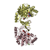
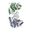
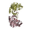
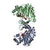
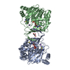
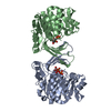
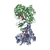
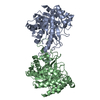
| Title | Structure of Burkholderia thailandensis nucleoside kinase (BthNK) in complex with inosine | ||||||
 Components Components | Ribokinase, putative | ||||||
 Keywords Keywords | TRANSFERASE / Rossmann Fold / kinase / ATP binding / Mg binding / Nucleoside binding | ||||||
| Function / homology |  Function and homology information Function and homology information | ||||||
| Biological species |  Burkholderia thailandensis (bacteria) Burkholderia thailandensis (bacteria) | ||||||
| Method |  X-RAY DIFFRACTION / SYNCHROTRON / MOLECULAR REPLACEMENT / Resolution: 1.7 Å X-RAY DIFFRACTION / SYNCHROTRON / MOLECULAR REPLACEMENT / Resolution: 1.7 Å | ||||||
 Authors Authors | Yasutake, Y. / Ota, H. / Hino, E. / Sakasegawa, S. / Tamura, T. | ||||||
 Citation Citation | Journal: Acta Crystallogr.,Sect.D / Year: 2011 Title: Structures of Burkholderia thailandensis nucleoside kinase: implications for the catalytic mechanism and nucleoside selectivity Authors: Yasutake, Y. / Ota, H. / Hino, E. / Sakasegawa, S. / Tamura, T. | ||||||
| History |
|
- Structure visualization
| Structure viewer | Molecule: MolmilJmol/JSmol |
|---|
- Downloads & links
-Download
| PDBx/mmCIF format | 3b1q.cif.gz | 723.2 KB | Display | PDBx/mmCIF format |
|---|---|---|---|---|
| PDB format | pdb3b1q.ent.gz | 597.2 KB | Display | PDB format |
| PDBx/mmJSON format | 3b1q.json.gz | Tree view | PDBx/mmJSON format | |
| Others |  Other downloads Other downloads |
-Validation report
| Arichive directory | https://data.pdbj.org/pub/pdb/validation_reports/b1/3b1qftp://data.pdbj.org/pub/pdb/validation_reports/b1/3b1q | HTTPS FTP |
|---|
-Related structure data
| Related structure data | 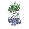 3b1nSC 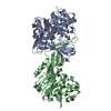 3b1oC 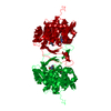 3b1pC 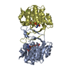 3b1rC S: Starting model for refinement C: citing same article ( |
|---|---|
| Similar structure data |


-Links
 PDBj
PDBj- Assembly
| Deposited unit | 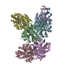
| ||||||||
|---|---|---|---|---|---|---|---|---|---|
| 1 | 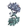
| ||||||||
| 2 | 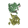
| ||||||||
| 3 | 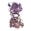
| ||||||||
| Unit cell |
|
-Components
-Protein , 1 types, 6 molecules ABCDEF
| #1: Protein | Mass: 35751.480 Da / Num. of mol.: 6 Source method: isolated from a genetically manipulated source Source: (gene. exp.) Burkholderia thailandensis (bacteria) / Strain: E264 / Gene: BTH_I1158 / Plasmid: pTip-QC2 / Production host: Rhodococcus erythropolis (bacteria) / Strain (production host): L88 / References: UniProt: Q2SZE4, EC: 2.7.1.143 |
|---|
-Non-polymers , 5 types, 1092 molecules 


| #2: Chemical | ChemComp-NOS /  Mass: 268.226 Da / Num. of mol.: 6 / Source method: obtained synthetically / Formula: C10H12N4O5 Mass: 268.226 Da / Num. of mol.: 6 / Source method: obtained synthetically / Formula: C10H12N4O5#3: Chemical | ChemComp-ACT /  Mass: 59.044 Da / Num. of mol.: 16 / Source method: obtained synthetically / Formula: C2H3O2 Mass: 59.044 Da / Num. of mol.: 16 / Source method: obtained synthetically / Formula: C2H3O2#4: Chemical | ChemComp-P33 /  Mass: 326.383 Da / Num. of mol.: 6 / Source method: obtained synthetically / Formula: C14H30O8 / Comment: precipitant*YM Mass: 326.383 Da / Num. of mol.: 6 / Source method: obtained synthetically / Formula: C14H30O8 / Comment: precipitant*YM#5: Chemical | ChemComp-NA /  Mass: 22.990 Da / Num. of mol.: 15 / Source method: obtained synthetically / Formula: Na Mass: 22.990 Da / Num. of mol.: 15 / Source method: obtained synthetically / Formula: Na#6: Water | ChemComp-HOH / | Mass: 18.015 Da / Num. of mol.: 1049 / Source method: isolated from a natural source / Formula: H2O |
|---|
-Experimental details
-Experiment
| Experiment | Method: X-RAY DIFFRACTION / Number of used crystals: 1 |
|---|
- Sample preparation
| Crystal | Density Matthews: 2.37 Å3/Da / Density % sol: 48.13 % |
|---|---|
| Crystal grow | Temperature: 293 K / Method: vapor diffusion / pH: 4 Details: 0.1M Na acetate, 0.2M Ca acetate, 26% PEG 400, pH 4.0, VAPOR DIFFUSION, temperature 293K |
-Data collection
| Diffraction | Mean temperature: 100 K |
|---|---|
| Diffraction source | Source: SYNCHROTRON / Site: Photon Factory  / Beamline: AR-NE3A / Wavelength: 1 Å / Beamline: AR-NE3A / Wavelength: 1 Å |
| Detector | Type: ADSC QUANTUM 270 / Detector: CCD / Date: Jun 21, 2009 |
| Radiation | Monochromator: Si(111) double crystal / Protocol: SINGLE WAVELENGTH / Monochromatic (M) / Laue (L): M / Scattering type: x-ray |
| Radiation wavelength | Wavelength: 1 Å / Relative weight: 1 |
| Reflection | Resolution: 1.7→50 Å / Num. obs: 208756 / % possible obs: 97.2 % / Redundancy: 3.9 % / Biso Wilson estimate: 14.8 Å2 / Rmerge(I) obs: 0.056 / Net I/σ(I): 19.1 |
| Reflection shell | Resolution: 1.7→1.73 Å / Redundancy: 3.6 % / Rmerge(I) obs: 0.376 / Mean I/σ(I) obs: 2.8 / % possible all: 94 |
- Processing
| Software |
| |||||||||||||||||||||||||||||||||||||||||||||||||||||||||||||||||||||||||||||||||||||||||||||||||||||||||||||||||||||||||||||||||||||||||||||||||||||||||||||||||||||||||||||||
|---|---|---|---|---|---|---|---|---|---|---|---|---|---|---|---|---|---|---|---|---|---|---|---|---|---|---|---|---|---|---|---|---|---|---|---|---|---|---|---|---|---|---|---|---|---|---|---|---|---|---|---|---|---|---|---|---|---|---|---|---|---|---|---|---|---|---|---|---|---|---|---|---|---|---|---|---|---|---|---|---|---|---|---|---|---|---|---|---|---|---|---|---|---|---|---|---|---|---|---|---|---|---|---|---|---|---|---|---|---|---|---|---|---|---|---|---|---|---|---|---|---|---|---|---|---|---|---|---|---|---|---|---|---|---|---|---|---|---|---|---|---|---|---|---|---|---|---|---|---|---|---|---|---|---|---|---|---|---|---|---|---|---|---|---|---|---|---|---|---|---|---|---|---|---|---|---|
| Refinement | Method to determine structure: MOLECULAR REPLACEMENT Starting model: PDB ENTRY 3B1N Resolution: 1.7→47.78 Å / Cor.coef. Fo:Fc: 0.961 / Cor.coef. Fo:Fc free: 0.948 / SU B: 3.774 / SU ML: 0.065 / Cross valid method: THROUGHOUT / ESU R Free: 0.1 / Stereochemistry target values: MAXIMUM LIKELIHOOD / Details: HYDROGENS HAVE BEEN ADDED IN THE RIDING POSITIONS
| |||||||||||||||||||||||||||||||||||||||||||||||||||||||||||||||||||||||||||||||||||||||||||||||||||||||||||||||||||||||||||||||||||||||||||||||||||||||||||||||||||||||||||||||
| Solvent computation | Ion probe radii: 0.8 Å / Shrinkage radii: 0.8 Å / VDW probe radii: 1.2 Å / Solvent model: MASK | |||||||||||||||||||||||||||||||||||||||||||||||||||||||||||||||||||||||||||||||||||||||||||||||||||||||||||||||||||||||||||||||||||||||||||||||||||||||||||||||||||||||||||||||
| Displacement parameters | Biso mean: 24.609 Å2
| |||||||||||||||||||||||||||||||||||||||||||||||||||||||||||||||||||||||||||||||||||||||||||||||||||||||||||||||||||||||||||||||||||||||||||||||||||||||||||||||||||||||||||||||
| Refinement step | Cycle: LAST / Resolution: 1.7→47.78 Å
| |||||||||||||||||||||||||||||||||||||||||||||||||||||||||||||||||||||||||||||||||||||||||||||||||||||||||||||||||||||||||||||||||||||||||||||||||||||||||||||||||||||||||||||||
| Refine LS restraints |
| |||||||||||||||||||||||||||||||||||||||||||||||||||||||||||||||||||||||||||||||||||||||||||||||||||||||||||||||||||||||||||||||||||||||||||||||||||||||||||||||||||||||||||||||
| LS refinement shell | Resolution: 1.704→1.748 Å / Total num. of bins used: 20
| |||||||||||||||||||||||||||||||||||||||||||||||||||||||||||||||||||||||||||||||||||||||||||||||||||||||||||||||||||||||||||||||||||||||||||||||||||||||||||||||||||||||||||||||
| Refinement TLS params. | Method: refined / Refine-ID: X-RAY DIFFRACTION
| |||||||||||||||||||||||||||||||||||||||||||||||||||||||||||||||||||||||||||||||||||||||||||||||||||||||||||||||||||||||||||||||||||||||||||||||||||||||||||||||||||||||||||||||
| Refinement TLS group |
|
