 Movie
Movie Controller

[English] 日本語
 Yorodumi
- PDB-4jg1: Structure of phosphoserine/threonine (pSTAb) scaffold bound to pT... -
+ Open data

- Basic information
| Entry | Database: PDB / ID: 4jg1 | ||||||
|---|---|---|---|---|---|---|---|
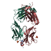
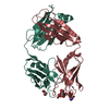
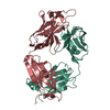
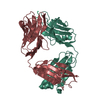
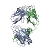
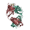
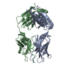
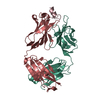
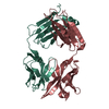
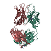
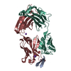
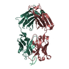
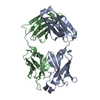
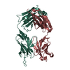
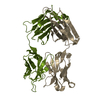
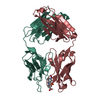
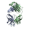
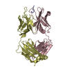
| Title | Structure of phosphoserine/threonine (pSTAb) scaffold bound to pThr peptide | ||||||
 Components Components |
| ||||||
 Keywords Keywords |  IMMUNE SYSTEM / immmunoglobulin domain IMMUNE SYSTEM / immmunoglobulin domain | ||||||
| Function / homology | Immunoglobulins / Immunoglobulin-like / Sandwich / Mainly Beta / PROPANOIC ACID Function and homology information Function and homology information | ||||||
| Biological species |  Homo sapiens (human) Homo sapiens (human) | ||||||
| Method | X-RAY DIFFRACTION / SYNCHROTRON / MOLECULAR REPLACEMENT / molecular replacement / Resolution: 1.55 Å | ||||||
 Authors Authors | Koerber, J.T. / Thomsen, N.D. / Hannigan, B.T. / Degrado, W.F. / Wells, J.A. | ||||||
 Citation Citation | Journal: Nat.Biotechnol. / Year: 2013 Title: Nature-inspired design of motif-specific antibody scaffolds. Authors: Koerber, J.T. / Thomsen, N.D. / Hannigan, B.T. / Degrado, W.F. / Wells, J.A. | ||||||
| History |
|
- Structure visualization
| Structure viewer | Molecule: MolmilJmol/JSmol |
|---|
- Downloads & links
-Download
| PDBx/mmCIF format | 4jg1.cif.gz | 281.5 KB | Display | PDBx/mmCIF format |
|---|---|---|---|---|
| PDB format | pdb4jg1.ent.gz | 230.7 KB | Display | PDB format |
| PDBx/mmJSON format | 4jg1.json.gz | Tree view | PDBx/mmJSON format | |
| Others |  Other downloads Other downloads |
-Validation report
| Arichive directory | https://data.pdbj.org/pub/pdb/validation_reports/jg/4jg1ftp://data.pdbj.org/pub/pdb/validation_reports/jg/4jg1 | HTTPS FTP |
|---|
-Related structure data
| Related structure data | 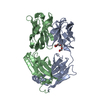 4jfxC 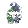 4jfyC 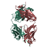 4jfzC 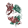 4jg0C 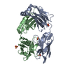 2gcyS 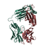 3n9gS C: citing same article ( S: Starting model for refinement |
|---|---|
| Similar structure data |


-Links
 PDBj
PDBj


- Assembly
| Deposited unit | 
| ||||||||
|---|---|---|---|---|---|---|---|---|---|
| 1 |
| ||||||||
| Unit cell |
| ||||||||
| Details | biological unit is the same as asym. |
-Components
| #1: Antibody | Fragment antigen-binding Mass: 23303.885 Da / Num. of mol.: 1 Source method: isolated from a genetically manipulated source Source: (gene. exp.) Homo sapiens (human) / Production host:  Escherichia coli (E. coli) / Strain (production host): C43 PRO+ Escherichia coli (E. coli) / Strain (production host): C43 PRO+ |
|---|---|
| #2: Antibody | Fragment antigen-binding Mass: 24790.717 Da / Num. of mol.: 1 Source method: isolated from a genetically manipulated source Source: (gene. exp.) Homo sapiens (human) / Production host: Escherichia coli (E. coli) / Strain (production host): C43 PRO+ |
| #3: Protein/peptide | Mass: 1415.465 Da / Num. of mol.: 1 / Source method: obtained synthetically |
| #4: Chemical | ChemComp-PPI / Propionic acid  Mass: 74.079 Da / Num. of mol.: 1 / Source method: obtained synthetically / Formula: C3H6O2 Mass: 74.079 Da / Num. of mol.: 1 / Source method: obtained synthetically / Formula: C3H6O2 |
| #5: Water | ChemComp-HOH / Water Mass: 18.015 Da / Num. of mol.: 621 / Source method: isolated from a natural source / Formula: H2O Mass: 18.015 Da / Num. of mol.: 621 / Source method: isolated from a natural source / Formula: H2O |
-Experimental details
-Experiment
| Experiment | Method: X-RAY DIFFRACTION / Number of used crystals: 1 |
|---|
- Sample preparation
| Crystal | Density Matthews: 2.53 Å3/Da / Density % sol: 51.45 % |
|---|---|
| Crystal grow | Temperature: 278 K / Method: vapor diffusion, hanging drop / pH: 6 Details: 25% PEG1500, 0.1M PCB, pH 6, VAPOR DIFFUSION, HANGING DROP, temperature 278K |
-Data collection
| Diffraction | Mean temperature: 100 K | |||||||||||||||||||||||||||||||||||||||||||||||||||||||||||||||||||||||||||||
|---|---|---|---|---|---|---|---|---|---|---|---|---|---|---|---|---|---|---|---|---|---|---|---|---|---|---|---|---|---|---|---|---|---|---|---|---|---|---|---|---|---|---|---|---|---|---|---|---|---|---|---|---|---|---|---|---|---|---|---|---|---|---|---|---|---|---|---|---|---|---|---|---|---|---|---|---|---|---|
| Diffraction source | Source: SYNCHROTRON / Site: ALS  / Beamline: 8.3.1 / Wavelength: 1.11587 Å / Beamline: 8.3.1 / Wavelength: 1.11587 Å | |||||||||||||||||||||||||||||||||||||||||||||||||||||||||||||||||||||||||||||
| Detector | Type: ADSC QUANTUM 315r / Detector: CCD / Date: Sep 5, 2012 | |||||||||||||||||||||||||||||||||||||||||||||||||||||||||||||||||||||||||||||
| Radiation | Monochromator: DOUBLE FLAT CRYSTAL, SI(111) / Protocol: SINGLE WAVELENGTH / Monochromatic (M) / Laue (L): M / Scattering type: x-ray | |||||||||||||||||||||||||||||||||||||||||||||||||||||||||||||||||||||||||||||
| Radiation wavelength | Wavelength: 1.11587 Å / Relative weight: 1 | |||||||||||||||||||||||||||||||||||||||||||||||||||||||||||||||||||||||||||||
| Reflection | Resolution: 1.55→50 Å / Num. all: 72575 / Num. obs: 72575 / % possible obs: 98 % / Observed criterion σ(F): 0 / Observed criterion σ(I): -3 / Redundancy: 5.6 % / Rmerge(I) obs: 0.065 / Χ2: 1.075 / Net I/σ(I): 13.5 | |||||||||||||||||||||||||||||||||||||||||||||||||||||||||||||||||||||||||||||
| Reflection shell |
|
-Phasing
| Phasing | Method: molecular replacement |
|---|
- Processing
| Software |
| |||||||||||||||||||||||||||||||||||||||||||||||||||||||||||||||||||||||||||||||||||||||||||||||||||||||||||||||||||||||||||||||||||||||||||||||||||||||||||||||||||||||||||||||||||||||||||||
|---|---|---|---|---|---|---|---|---|---|---|---|---|---|---|---|---|---|---|---|---|---|---|---|---|---|---|---|---|---|---|---|---|---|---|---|---|---|---|---|---|---|---|---|---|---|---|---|---|---|---|---|---|---|---|---|---|---|---|---|---|---|---|---|---|---|---|---|---|---|---|---|---|---|---|---|---|---|---|---|---|---|---|---|---|---|---|---|---|---|---|---|---|---|---|---|---|---|---|---|---|---|---|---|---|---|---|---|---|---|---|---|---|---|---|---|---|---|---|---|---|---|---|---|---|---|---|---|---|---|---|---|---|---|---|---|---|---|---|---|---|---|---|---|---|---|---|---|---|---|---|---|---|---|---|---|---|---|---|---|---|---|---|---|---|---|---|---|---|---|---|---|---|---|---|---|---|---|---|---|---|---|---|---|---|---|---|---|---|---|---|
| Refinement | Method to determine structure: MOLECULAR REPLACEMENT Starting model: pdb entries 3N9G, 2GCY Resolution: 1.55→41.143 Å / Occupancy max: 1 / Occupancy min: 0.08 / FOM work R set: 0.9044 / SU ML: 0.11 / σ(F): 1.36 / Phase error: 16.07 / Stereochemistry target values: ML
| |||||||||||||||||||||||||||||||||||||||||||||||||||||||||||||||||||||||||||||||||||||||||||||||||||||||||||||||||||||||||||||||||||||||||||||||||||||||||||||||||||||||||||||||||||||||||||||
| Solvent computation | Shrinkage radii: 0.9 Å / VDW probe radii: 1.11 Å / Solvent model: FLAT BULK SOLVENT MODEL | |||||||||||||||||||||||||||||||||||||||||||||||||||||||||||||||||||||||||||||||||||||||||||||||||||||||||||||||||||||||||||||||||||||||||||||||||||||||||||||||||||||||||||||||||||||||||||||
| Displacement parameters | Biso max: 93.37 Å2 / Biso mean: 21.011 Å2 / Biso min: 8.14 Å2 | |||||||||||||||||||||||||||||||||||||||||||||||||||||||||||||||||||||||||||||||||||||||||||||||||||||||||||||||||||||||||||||||||||||||||||||||||||||||||||||||||||||||||||||||||||||||||||||
| Refinement step | Cycle: LAST / Resolution: 1.55→41.143 Å
| |||||||||||||||||||||||||||||||||||||||||||||||||||||||||||||||||||||||||||||||||||||||||||||||||||||||||||||||||||||||||||||||||||||||||||||||||||||||||||||||||||||||||||||||||||||||||||||
| Refine LS restraints |
| |||||||||||||||||||||||||||||||||||||||||||||||||||||||||||||||||||||||||||||||||||||||||||||||||||||||||||||||||||||||||||||||||||||||||||||||||||||||||||||||||||||||||||||||||||||||||||||
| LS refinement shell | Refine-ID: X-RAY DIFFRACTION / Total num. of bins used: 26
| |||||||||||||||||||||||||||||||||||||||||||||||||||||||||||||||||||||||||||||||||||||||||||||||||||||||||||||||||||||||||||||||||||||||||||||||||||||||||||||||||||||||||||||||||||||||||||||
| Refinement TLS params. | Method: refined / Refine-ID: X-RAY DIFFRACTION
| |||||||||||||||||||||||||||||||||||||||||||||||||||||||||||||||||||||||||||||||||||||||||||||||||||||||||||||||||||||||||||||||||||||||||||||||||||||||||||||||||||||||||||||||||||||||||||||
| Refinement TLS group |
|
