 Movie
Movie Controller

[English] 日本語
 Yorodumi
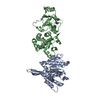
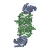
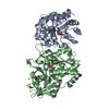
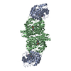
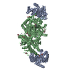
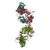
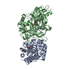
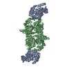
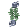
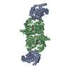
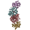
- PDB-4ilg: Crystal structure of Aar2p in complex with the Prp8p RNaseH and J... -
+ Open data

- Basic information
| Entry | Database: PDB / ID: 4ilg | ||||||
|---|---|---|---|---|---|---|---|
| Title | Crystal structure of Aar2p in complex with the Prp8p RNaseH and Jab1/MPN domains | ||||||
 Components Components |
| ||||||
 Keywords Keywords |  SPLICING / U5 snRNP assembly / Aar2 / Prp8 SPLICING / U5 snRNP assembly / Aar2 / Prp8 | ||||||
| Function / homology |  Function and homology information Function and homology informationgeneration of catalytic spliceosome for second transesterification step / mRNA 3'-splice site recognition / mRNA 5'-splice site recognition / spliceosomal tri-snRNP complex assembly / U5 snRNA binding / U5 snRNP / U2 snRNA binding / U6 snRNA binding / spliceosomal snRNP assembly / pre-mRNA intronic binding ...generation of catalytic spliceosome for second transesterification step / mRNA 3'-splice site recognition / mRNA 5'-splice site recognition / spliceosomal tri-snRNP complex assembly / U5 snRNA binding / U5 snRNP / U2 snRNA binding / U6 snRNA binding / spliceosomal snRNP assembly / pre-mRNA intronic binding / U1 snRNA binding / U4/U6 x U5 tri-snRNP complex / catalytic step 2 spliceosome / spliceosomal complex / mRNA splicing, via spliceosome / metallopeptidase activity / mRNA binding / nucleus / cytoplasmSimilarity search - Function | ||||||
| Biological species |  Saccharomyces cerevisiae (brewer's yeast) Saccharomyces cerevisiae (brewer's yeast) | ||||||
| Method | X-RAY DIFFRACTION / SYNCHROTRON / MOLECULAR REPLACEMENT / Resolution: 2.1 Å | ||||||
 Authors Authors | Weber, G. / Heroven, A.C. / Santos, K.F. / Wahl, M.C. | ||||||
 Citation Citation | Journal: Genes Dev. / Year: 2013 Title: Structural basis for dual roles of Aar2p in U5 snRNP assembly. Authors: Weber, G. / Cristao, V.F. / Santos, K.F. / Jovin, S.M. / Heroven, A.C. / Holton, N. / Luhrmann, R. / Beggs, J.D. / Wahl, M.C. | ||||||
| History |
|
- Structure visualization
| Structure viewer | Molecule: MolmilJmol/JSmol |
|---|
- Downloads & links
-Download
| PDBx/mmCIF format | 4ilg.cif.gz | 370 KB | Display | PDBx/mmCIF format |
|---|---|---|---|---|
| PDB format | pdb4ilg.ent.gz | 299.2 KB | Display | PDB format |
| PDBx/mmJSON format | 4ilg.json.gz | Tree view | PDBx/mmJSON format | |
| Others |  Other downloads Other downloads |
-Validation report
| Arichive directory | https://data.pdbj.org/pub/pdb/validation_reports/il/4ilgftp://data.pdbj.org/pub/pdb/validation_reports/il/4ilg | HTTPS FTP |
|---|
-Related structure data
| Related structure data | 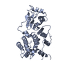 4iliC 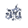 4iljC 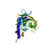 2og4S 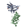 3sbtS C: citing same article ( S: Starting model for refinement |
|---|---|
| Similar structure data |
-Links
 PDBj
PDBj

- Assembly
| Deposited unit | 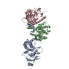
| ||||||||
|---|---|---|---|---|---|---|---|---|---|
| 1 |
| ||||||||
| Unit cell |
|
-Components
| #1: Protein | Mass: 40219.867 Da / Num. of mol.: 1 / Mutation: Residues 153-170 replaced by 5 Serines Source method: isolated from a genetically manipulated source Source: (gene. exp.) Saccharomyces cerevisiae (brewer's yeast)Strain: ATCC 204508 / S288c / Gene: AAR2, YBL074C, YBL06.06, YBL0611 / Production host:  Escherichia coli (E. coli) / References: UniProt: P32357 Escherichia coli (E. coli) / References: UniProt: P32357 |
|---|---|
| #2: Protein | Mass: 29501.113 Da / Num. of mol.: 1 / Fragment: yPrp8 RNaseH (UNP Residues 1835-2090) Source method: isolated from a genetically manipulated source Source: (gene. exp.) Saccharomyces cerevisiae (brewer's yeast)Strain: ATCC 204508 / S288c / Gene: PRP8, DBF3, DNA39, RNA8, SLT21, USA2, YHR165C / Production host: Escherichia coli (E. coli) / References: UniProt: P33334 |
| #3: Protein | Mass: 30490.338 Da / Num. of mol.: 1 / Fragment: yPrp8 Jab1/MPN (UNP Residues 2147-2413) Source method: isolated from a genetically manipulated source Source: (gene. exp.) Saccharomyces cerevisiae (brewer's yeast)Strain: ATCC 204508 / S288c / Gene: PRP8, DBF3, DNA39, RNA8, SLT21, USA2, YHR165C / Production host: Escherichia coli (E. coli) / References: UniProt: P33334 |
| #4: Chemical | ChemComp-EPE / HEPES  Mass: 238.305 Da / Num. of mol.: 1 / Source method: obtained synthetically / Formula: C8H18N2O4S / Comment: pH buffer*YM Mass: 238.305 Da / Num. of mol.: 1 / Source method: obtained synthetically / Formula: C8H18N2O4S / Comment: pH buffer*YM |
| #5: Water | ChemComp-HOH / Water Mass: 18.015 Da / Num. of mol.: 689 / Source method: isolated from a natural source / Formula: H2O Mass: 18.015 Da / Num. of mol.: 689 / Source method: isolated from a natural source / Formula: H2O |
-Experimental details
-Experiment
| Experiment | Method: X-RAY DIFFRACTION / Number of used crystals: 1 |
|---|
- Sample preparation
| Crystal | Density Matthews: 2.97 Å3/Da / Density % sol: 58.53 % |
|---|---|
| Crystal grow | Temperature: 277 K / Method: vapor diffusion, sitting drop / pH: 7.5 Details: 0.1 M HEPES-NaOH, pH 7.5, 12 % (w/v) PEG 6000 and 0.6 M KCl, VAPOR DIFFUSION, SITTING DROP, temperature 277K |
-Data collection
| Diffraction | Mean temperature: 100 K |
|---|---|
| Diffraction source | Source: SYNCHROTRON / Site: BESSY  / Beamline: 14.2 / Wavelength: 0.9184 Å / Beamline: 14.2 / Wavelength: 0.9184 Å |
| Detector | Type: RAYONIX MX-225 / Detector: CCD / Date: Nov 30, 2011 |
| Radiation | Monochromator: Si-111 crystal / Protocol: SINGLE WAVELENGTH / Monochromatic (M) / Laue (L): M / Scattering type: x-ray |
| Radiation wavelength | Wavelength: 0.9184 Å / Relative weight: 1 |
| Reflection | Resolution: 2.1→34.3 Å / Num. all: 68910 / Num. obs: 68679 / % possible obs: 99.9 % / Observed criterion σ(F): 1.6 / Observed criterion σ(I): 1.6 / Redundancy: 3.4 % / Rmerge(I) obs: 0.207 / Rsym value: 0.09 / Net I/σ(I): 10.6 |
| Reflection shell | Resolution: 2.1→2.15 Å / % possible obs: 99.9 % / Redundancy: 3.5 % / Rmerge(I) obs: 1.154 / Mean I/σ(I) obs: 1.6 / Rsym value: 0.861 |
- Processing
| Software |
| |||||||||||||||||||||||||||||||||||||||||||||||||||||||||||||||||||||||||||||||||||||||||||||||||||||||||||||||||||||||||||||||||||||||||||||||||||||||||||||||||
|---|---|---|---|---|---|---|---|---|---|---|---|---|---|---|---|---|---|---|---|---|---|---|---|---|---|---|---|---|---|---|---|---|---|---|---|---|---|---|---|---|---|---|---|---|---|---|---|---|---|---|---|---|---|---|---|---|---|---|---|---|---|---|---|---|---|---|---|---|---|---|---|---|---|---|---|---|---|---|---|---|---|---|---|---|---|---|---|---|---|---|---|---|---|---|---|---|---|---|---|---|---|---|---|---|---|---|---|---|---|---|---|---|---|---|---|---|---|---|---|---|---|---|---|---|---|---|---|---|---|---|---|---|---|---|---|---|---|---|---|---|---|---|---|---|---|---|---|---|---|---|---|---|---|---|---|---|---|---|---|---|---|---|
| Refinement | Method to determine structure: MOLECULAR REPLACEMENT Starting model: PDB ENTRY 3SBT, 2OG4 Resolution: 2.1→31.869 Å / SU ML: 0.29 / σ(F): 1.99 / Phase error: 23.77 / Stereochemistry target values: ML
| |||||||||||||||||||||||||||||||||||||||||||||||||||||||||||||||||||||||||||||||||||||||||||||||||||||||||||||||||||||||||||||||||||||||||||||||||||||||||||||||||
| Solvent computation | Shrinkage radii: 0.98 Å / VDW probe radii: 1.2 Å / Solvent model: FLAT BULK SOLVENT MODEL / Bsol: 46.555 Å2 / ksol: 0.291 e/Å3 | |||||||||||||||||||||||||||||||||||||||||||||||||||||||||||||||||||||||||||||||||||||||||||||||||||||||||||||||||||||||||||||||||||||||||||||||||||||||||||||||||
| Displacement parameters |
| |||||||||||||||||||||||||||||||||||||||||||||||||||||||||||||||||||||||||||||||||||||||||||||||||||||||||||||||||||||||||||||||||||||||||||||||||||||||||||||||||
| Refinement step | Cycle: LAST / Resolution: 2.1→31.869 Å
| |||||||||||||||||||||||||||||||||||||||||||||||||||||||||||||||||||||||||||||||||||||||||||||||||||||||||||||||||||||||||||||||||||||||||||||||||||||||||||||||||
| Refine LS restraints |
| |||||||||||||||||||||||||||||||||||||||||||||||||||||||||||||||||||||||||||||||||||||||||||||||||||||||||||||||||||||||||||||||||||||||||||||||||||||||||||||||||
| LS refinement shell |
| |||||||||||||||||||||||||||||||||||||||||||||||||||||||||||||||||||||||||||||||||||||||||||||||||||||||||||||||||||||||||||||||||||||||||||||||||||||||||||||||||
| Refinement TLS params. | Method: refined / Refine-ID: X-RAY DIFFRACTION
| |||||||||||||||||||||||||||||||||||||||||||||||||||||||||||||||||||||||||||||||||||||||||||||||||||||||||||||||||||||||||||||||||||||||||||||||||||||||||||||||||
| Refinement TLS group |
|
