 Movie
Movie Controller


+ Open data

- Basic information
| Entry | Database: PDB / ID: 7ndi | ||||||
|---|---|---|---|---|---|---|---|
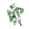
| Title | Crystal structure of ZC3H12C PIN domain with Mg2+ Ion | ||||||
 Components Components | Probable ribonuclease ZC3H12C | ||||||
 Keywords Keywords | RNA BINDING PROTEIN / Endo-ribonuclease / Regnase | ||||||
| Function / homology |  Function and homology information Function and homology informationRNA endonuclease activity / cytoplasmic ribonucleoprotein granule / Hydrolases; Acting on ester bonds / hydrolase activity / mRNA binding / zinc ion binding / nucleus Similarity search - Function | ||||||
| Biological species |  | ||||||
| Method |  X-RAY DIFFRACTION / SYNCHROTRON / MOLECULAR REPLACEMENT / Resolution: 2.875 Å X-RAY DIFFRACTION / SYNCHROTRON / MOLECULAR REPLACEMENT / Resolution: 2.875 Å | ||||||
 Authors Authors | Garg, A. / Heinemann, U. | ||||||
 Citation Citation | Journal: Nucleic Acids Res. / Year: 2021 Title: PIN and CCCH Zn-finger domains coordinate RNA targeting in ZC3H12 family endoribonucleases. Authors: Garg, A. / Roske, Y. / Yamada, S. / Uehata, T. / Takeuchi, O. / Heinemann, U. | ||||||
| History |
|
- Structure visualization
| Structure viewer | Molecule: MolmilJmol/JSmol |
|---|
- Downloads & links
-Download
| PDBx/mmCIF format | 7ndi.cif.gz | 293.5 KB | Display | PDBx/mmCIF format |
|---|---|---|---|---|
| PDB format | pdb7ndi.ent.gz | 240.6 KB | Display | PDB format |
| PDBx/mmJSON format | 7ndi.json.gz | Tree view | PDBx/mmJSON format | |
| Others |  Other downloads Other downloads |
-Validation report
| Arichive directory | https://data.pdbj.org/pub/pdb/validation_reports/nd/7ndiftp://data.pdbj.org/pub/pdb/validation_reports/nd/7ndi | HTTPS FTP |
|---|
-Related structure data
| Related structure data | 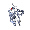 7ndhC 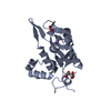 7ndjC 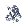 7ndkC  3v34S S: Starting model for refinement C: citing same article ( |
|---|---|
| Similar structure data |


-Links
 PDBj
PDBj
- Assembly
| Deposited unit | 
| |||||||||||||||||||||||||||||||||||||||||||||||||||||||
|---|---|---|---|---|---|---|---|---|---|---|---|---|---|---|---|---|---|---|---|---|---|---|---|---|---|---|---|---|---|---|---|---|---|---|---|---|---|---|---|---|---|---|---|---|---|---|---|---|---|---|---|---|---|---|---|---|
| 1 | 
| |||||||||||||||||||||||||||||||||||||||||||||||||||||||
| 2 | 
| |||||||||||||||||||||||||||||||||||||||||||||||||||||||
| 3 | 
| |||||||||||||||||||||||||||||||||||||||||||||||||||||||
| 4 | 
| |||||||||||||||||||||||||||||||||||||||||||||||||||||||
| Unit cell |
| |||||||||||||||||||||||||||||||||||||||||||||||||||||||
| Noncrystallographic symmetry (NCS) | NCS domain:
NCS domain segments: Component-ID: 1 / Ens-ID: 1 / End auth comp-ID: LYS / End label comp-ID: LYS
|
-Components
| #1: Protein | Mass: 19282.117 Da / Num. of mol.: 4 Source method: isolated from a genetically manipulated source Source: (gene. exp.)  References: UniProt: Q5DTV4, Hydrolases; Acting on ester bonds #2: Chemical | ChemComp-MG /   Mass: 24.305 Da / Num. of mol.: 4 / Source method: obtained synthetically / Formula: Mg / Feature type: SUBJECT OF INVESTIGATION Mass: 24.305 Da / Num. of mol.: 4 / Source method: obtained synthetically / Formula: Mg / Feature type: SUBJECT OF INVESTIGATION#3: Chemical |   Mass: 22.990 Da / Num. of mol.: 3 / Source method: obtained synthetically / Formula: Na Mass: 22.990 Da / Num. of mol.: 3 / Source method: obtained synthetically / Formula: Na#4: Chemical |   Mass: 62.068 Da / Num. of mol.: 3 / Source method: obtained synthetically / Formula: C2H6O2 Mass: 62.068 Da / Num. of mol.: 3 / Source method: obtained synthetically / Formula: C2H6O2#5: Water | ChemComp-HOH / |  Mass: 18.015 Da / Num. of mol.: 205 / Source method: isolated from a natural source / Formula: H2O Mass: 18.015 Da / Num. of mol.: 205 / Source method: isolated from a natural source / Formula: H2OHas ligand of interest | Y | |
|---|
-Experimental details
-Experiment
| Experiment | Method: X-RAY DIFFRACTION / Number of used crystals: 1 |
|---|
- Sample preparation
| Crystal | Density Matthews: 6.34 Å3/Da / Density % sol: 80.58 % |
|---|---|
| Crystal grow | Temperature: 293 K / Method: vapor diffusion, sitting drop / Details: 0.2 M LiCl, 50 mM MgSO4 and 8% PEG 8000 |
-Data collection
| Diffraction | Mean temperature: 100 K / Serial crystal experiment: N |
|---|---|
| Diffraction source | Source: SYNCHROTRON / Site: BESSY  / Beamline: 14.1 / Wavelength: 0.91841 Å / Beamline: 14.1 / Wavelength: 0.91841 Å |
| Detector | Type: DECTRIS PILATUS3 S 6M / Detector: PIXEL / Date: Jul 19, 2015 |
| Radiation | Protocol: SINGLE WAVELENGTH / Monochromatic (M) / Laue (L): M / Scattering type: x-ray |
| Radiation wavelength | Wavelength: 0.91841 Å / Relative weight: 1 |
| Reflection | Resolution: 2.87→48.77 Å / Num. obs: 45274 / % possible obs: 99.53 % / Redundancy: 5.77 % / CC1/2: 0.997 / Net I/σ(I): 13.44 |
| Reflection shell | Resolution: 2.875→2.978 Å / Num. unique obs: 4314 / CC1/2: 0.717 |
- Processing
| Software |
| ||||||||||||||||||||||||||||||||||||||||||||||||||||||||||||||||||||||||||||||||||||||||||||||||
|---|---|---|---|---|---|---|---|---|---|---|---|---|---|---|---|---|---|---|---|---|---|---|---|---|---|---|---|---|---|---|---|---|---|---|---|---|---|---|---|---|---|---|---|---|---|---|---|---|---|---|---|---|---|---|---|---|---|---|---|---|---|---|---|---|---|---|---|---|---|---|---|---|---|---|---|---|---|---|---|---|---|---|---|---|---|---|---|---|---|---|---|---|---|---|---|---|---|
| Refinement | Method to determine structure: MOLECULAR REPLACEMENT Starting model: 3v34 Resolution: 2.875→48.767 Å / SU ML: 0.36 / Cross valid method: FREE R-VALUE / σ(F): 1.35 / Phase error: 23.77 / Stereochemistry target values: ML
| ||||||||||||||||||||||||||||||||||||||||||||||||||||||||||||||||||||||||||||||||||||||||||||||||
| Solvent computation | Shrinkage radii: 0.9 Å / VDW probe radii: 1.11 Å / Solvent model: FLAT BULK SOLVENT MODEL | ||||||||||||||||||||||||||||||||||||||||||||||||||||||||||||||||||||||||||||||||||||||||||||||||
| Displacement parameters | Biso max: 180.65 Å2 / Biso mean: 59.82 Å2 / Biso min: 44.61 Å2 | ||||||||||||||||||||||||||||||||||||||||||||||||||||||||||||||||||||||||||||||||||||||||||||||||
| Refinement step | Cycle: final / Resolution: 2.875→48.767 Å
| ||||||||||||||||||||||||||||||||||||||||||||||||||||||||||||||||||||||||||||||||||||||||||||||||
| Refine LS restraints |
| ||||||||||||||||||||||||||||||||||||||||||||||||||||||||||||||||||||||||||||||||||||||||||||||||
| Refine LS restraints NCS |
| ||||||||||||||||||||||||||||||||||||||||||||||||||||||||||||||||||||||||||||||||||||||||||||||||
| LS refinement shell | Refine-ID: X-RAY DIFFRACTION / Rfactor Rfree error: 0
| ||||||||||||||||||||||||||||||||||||||||||||||||||||||||||||||||||||||||||||||||||||||||||||||||
| Refinement TLS params. | Method: refined / Origin x: 107.1049 Å / Origin y: 85.0175 Å / Origin z: 21.2785 Å
| ||||||||||||||||||||||||||||||||||||||||||||||||||||||||||||||||||||||||||||||||||||||||||||||||
| Refinement TLS group |
|
