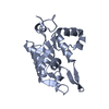
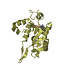
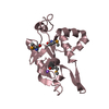
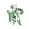
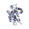
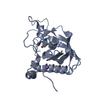
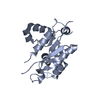
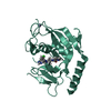
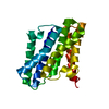
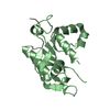
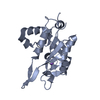
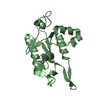
[English] 日本語
Yorodumi
- PDB-5h9w: Crystal structure of Regnase PIN domain, form II -

+
Open data


ID or keywords:

Loading...

-
Basic information

Entry
Database: PDB / ID: 5h9w
TitleCrystal structure of Regnase PIN domain, form II
ComponentsRibonuclease ZC3H12A
KeywordsHYDROLASE / RNase
Function / homology
Function and homology information


immune response-activating signaling pathway / positive regulation of miRNA catabolic process / positive regulation of protein deubiquitination / cellular response to ionomycin / miRNA catabolic process / RNA exonuclease activity / positive regulation of mRNA catabolic process / rough endoplasmic reticulum membrane / negative regulation by host of viral genome replication / cellular response to sodium arsenite ...immune response-activating signaling pathway / positive regulation of miRNA catabolic process / positive regulation of protein deubiquitination / cellular response to ionomycin / miRNA catabolic process / RNA exonuclease activity / positive regulation of mRNA catabolic process / rough endoplasmic reticulum membrane / negative regulation by host of viral genome replication / cellular response to sodium arsenite / negative regulation of T-helper 17 cell differentiation / negative regulation of muscle cell apoptotic process / nuclease activity / 3'-UTR-mediated mRNA destabilization / positive regulation of lipid storage / negative regulation of cardiac muscle contraction / negative regulation of nitric oxide biosynthetic process / mRNA 3'-UTR AU-rich region binding / cellular response to chemokine / negative regulation of non-canonical NF-kappaB signal transduction / nuclear-transcribed mRNA catabolic process, nonsense-mediated decay / miRNA binding / negative regulation of interleukin-1 beta production / positive regulation of p38MAPK cascade / negative regulation of NF-kappaB transcription factor activity / protein deubiquitination / positive regulation of execution phase of apoptosis / mRNA catabolic process / negative regulation of interleukin-6 production / negative regulation of type II interferon production / negative regulation of tumor necrosis factor production / negative regulation of cytokine production involved in inflammatory response / positive regulation of fat cell differentiation / cellular response to interleukin-1 / cellular response to glucose starvation / positive regulation of autophagy / negative regulation of canonical NF-kappaB signal transduction / rough endoplasmic reticulum / positive regulation of defense response to virus by host / RNA endonuclease activity / positive regulation of endothelial cell migration / negative regulation of protein phosphorylation / mRNA 3'-UTR binding / P-body / cellular response to virus / cytoplasmic ribonucleoprotein granule / positive regulation of protein import into nucleus / RNA stem-loop binding / positive regulation of angiogenesis / positive regulation of reactive oxygen species metabolic process / ribosome binding / protein complex oligomerization / cellular response to tumor necrosis factor / nervous system development / T cell receptor signaling pathway / cellular response to oxidative stress / cellular response to lipopolysaccharide / angiogenesis / cysteine-type deubiquitinase activity / Hydrolases; Acting on ester bonds / cell differentiation / cytoskeleton / inflammatory response / negative regulation of gene expression / mRNA binding / apoptotic process / DNA damage response / chromatin binding / positive regulation of gene expression / positive regulation of transcription by RNA polymerase II / protein-containing complex / DNA binding / nucleoplasm / nucleus / metal ion binding / cytoplasm
Similarity search - Function
Rege-1, UBA-like domain / Endoribonuclease Regnase 1/ZC3H12, C-terminal domain / UBA-like domain / Endoribonuclease Regnase 1/ ZC3H12 C-terminal domain / Rossmann fold - #11980 / Ribonuclease Zc3h12a-like, NYN domain / Zc3h12a-like Ribonuclease NYN domain / Rossmann fold / 3-Layer(aba) Sandwich / Alpha Beta
Similarity search - Domain/homology
Endoribonuclease ZC3H12A
Similarity search - Component
Biological speciesMus musculus (house mouse)
MethodX-RAY DIFFRACTION / SYNCHROTRON / MOLECULAR REPLACEMENT / Resolution: 2.6 Å
AuthorsYokogawa, M. / Tsushima, T. / Adachi, W. / Noda, N.N. / Inagaki, F.
Funding support Japan, 1items
OrganizationGrant numberCountry
JSPS Japan
CitationJournal: Sci Rep / Year: 2016
Title: Structural basis for the regulation of enzymatic activity of Regnase-1 by domain-domain interactions
Authors: Yokogawa, M. / Tsushima, T. / Noda, N.N. / Kumeta, H. / Enokizono, Y. / Yamashita, K. / Standley, D.M. / Takeuchi, O. / Akira, S. / Inagaki, F.
History
DepositionDec 29, 2015Deposition site: RCSB / Processing site: PDBJ
Revision 1.0Mar 16, 2016Provider: repository / Type: Initial release
Revision 1.1Feb 19, 2020Group: Data collection / Derived calculations / Category: diffrn_source / pdbx_struct_oper_list
Item: _diffrn_source.pdbx_synchrotron_site / _pdbx_struct_oper_list.symmetry_operation
Revision 1.2Nov 8, 2023Group: Data collection / Database references / Refinement description
Category: chem_comp_atom / chem_comp_bond ...chem_comp_atom / chem_comp_bond / database_2 / pdbx_initial_refinement_model / struct_ncs_dom_lim
Item: _database_2.pdbx_DOI / _database_2.pdbx_database_accession ..._database_2.pdbx_DOI / _database_2.pdbx_database_accession / _struct_ncs_dom_lim.beg_auth_comp_id / _struct_ncs_dom_lim.beg_label_asym_id / _struct_ncs_dom_lim.beg_label_comp_id / _struct_ncs_dom_lim.beg_label_seq_id / _struct_ncs_dom_lim.end_auth_comp_id / _struct_ncs_dom_lim.end_label_asym_id / _struct_ncs_dom_lim.end_label_comp_id / _struct_ncs_dom_lim.end_label_seq_id

-
Structure visualization

Structure viewerMolecule:
MolmilJmol/JSmol

Downloads & links

-
Assembly

Deposited unit
A: Ribonuclease ZC3H12A
B: Ribonuclease ZC3H12A
hetero molecules


Theoretical massNumber of molelcules
Total (without water)49,1074
Polymers49,0612
Non-polymers462
Water61334
1
A: Ribonuclease ZC3H12A
hetero molecules


Theoretical massNumber of molelcules
Total (without water)24,5532
Polymers24,5301
Non-polymers231
Water181
TypeNameSymmetry operationNumber
identity operation1_555x,y,z1
2
B: Ribonuclease ZC3H12A
hetero molecules


Theoretical massNumber of molelcules
Total (without water)24,5532
Polymers24,5301
Non-polymers231
Water181
TypeNameSymmetry operationNumber
identity operation1_555x,y,z1
Unit cell
Length a, b, c (Å)113.333, 113.333, 78.373
Angle α, β, γ (deg.)90.00, 90.00, 120.00
Int Tables number152
Space group name H-MP3121
Noncrystallographic symmetry (NCS)NCS domain:
IDEns-IDDetails
11A
21B

NCS domain segments:

Component-ID: _ / Ens-ID: 1 / Beg auth comp-ID: ASP / Beg label comp-ID: ASP / End auth comp-ID: LYS / End label comp-ID: LYS / Refine code: _ / Auth seq-ID: 134 - 295 / Label seq-ID: 5 - 166

Dom-IDAuth asym-IDLabel asym-ID
1AA
2BB

-
Components

#1: Protein Ribonuclease ZC3H12A / Regnase-1 / MCP-induced protein 1 / Zinc finger CCCH domain-containing protein 12A


Mass: 24530.289 Da / Num. of mol.: 2 / Fragment: PIN domain, UNP residues 134-339
Source method: isolated from a genetically manipulated source
Source: (gene. exp.) Mus musculus (house mouse) / Gene: Zc3h12a, Mcpip, Mcpip1 / Production host: Escherichia coli (E. coli)
References: UniProt: Q5D1E7, Hydrolases; Acting on ester bonds
#2: Chemical ChemComp-NA / SODIUM ION


Mass: 22.990 Da / Num. of mol.: 2 / Source method: obtained synthetically / Formula: Na
#3: Water ChemComp-HOH / water


Mass: 18.015 Da / Num. of mol.: 34 / Source method: isolated from a natural source / Formula: H2O

-
Experimental details

-
Experiment

ExperimentMethod: X-RAY DIFFRACTION

-
Sample preparation

CrystalDensity Matthews: 2.96 Å3/Da / Density % sol: 58.47 %
Crystal growTemperature: 293 K / Method: vapor diffusion, sitting drop / pH: 7 / Details: 1.7 M NaCl, 100 mM HEPES

-
Data collection

DiffractionMean temperature: 95 K
Diffraction sourceSource: SYNCHROTRON / Site: SPring-8 / Beamline: BL41XU / Wavelength: 1 Å
DetectorType: ADSC QUANTUM 315 / Detector: CCD / Date: Apr 27, 2009
RadiationProtocol: SINGLE WAVELENGTH / Monochromatic (M) / Laue (L): M / Scattering type: x-ray
Radiation wavelengthWavelength: 1 Å / Relative weight: 1
ReflectionResolution: 2.6→26.94 Å / Num. obs: 16940 / % possible obs: 92.9 % / Redundancy: 10.4 % / Net I/σ(I): 34.3
Reflection shellResolution: 2.6→2.69 Å / Redundancy: 8.2 % / Rmerge(I) obs: 0.255 / Mean I/σ(I) obs: 7.9 / % possible all: 67.8

-
Processing

Software
NameVersionClassification
REFMAC5.7.0032refinement
HKL-2000data reduction
HKL-2000data scaling
CNSphasing
RefinementMethod to determine structure: MOLECULAR REPLACEMENT
Starting model: 5H9V
Resolution: 2.6→26.94 Å / Cor.coef. Fo:Fc: 0.958 / Cor.coef. Fo:Fc free: 0.935 / SU B: 20.815 / SU ML: 0.198 / Cross valid method: THROUGHOUT / ESU R: 0.371 / ESU R Free: 0.257 / Stereochemistry target values: MAXIMUM LIKELIHOOD / Details: HYDROGENS HAVE BEEN ADDED IN THE RIDING POSITIONS
RfactorNum. reflection% reflectionSelection details
Rfree0.23142 862 5.1 %RANDOM
Rwork0.19144 ---
obs0.19347 16057 92.67 %-
Solvent computationIon probe radii: 0.8 Å / Shrinkage radii: 0.8 Å / VDW probe radii: 1.2 Å / Solvent model: MASK
Displacement parametersBiso mean: 73.875 Å2
Baniso -1Baniso -2Baniso -3
1--3.21 Å2-3.21 Å2-0 Å2
2---3.21 Å2-0 Å2
3---10.41 Å2
Refinement stepCycle: 1 / Resolution: 2.6→26.94 Å
ProteinNucleic acidLigandSolventTotal
Num. atoms2658 0 2 34 2694
Refine LS restraints
Refine-IDTypeDev idealDev ideal targetNumber
X-RAY DIFFRACTIONr_bond_refined_d0.0150.0192722
X-RAY DIFFRACTIONr_bond_other_d0.0050.022632
X-RAY DIFFRACTIONr_angle_refined_deg1.7181.9583677
X-RAY DIFFRACTIONr_angle_other_deg1.15536045
X-RAY DIFFRACTIONr_dihedral_angle_1_deg6.7785322
X-RAY DIFFRACTIONr_dihedral_angle_2_deg35.65122.662139
X-RAY DIFFRACTIONr_dihedral_angle_3_deg19.72215485
X-RAY DIFFRACTIONr_dihedral_angle_4_deg18.4951531
X-RAY DIFFRACTIONr_chiral_restr0.090.2392
X-RAY DIFFRACTIONr_gen_planes_refined0.010.0213037
X-RAY DIFFRACTIONr_gen_planes_other0.0050.02658
X-RAY DIFFRACTIONr_nbd_refined
X-RAY DIFFRACTIONr_nbd_other
X-RAY DIFFRACTIONr_nbtor_refined
X-RAY DIFFRACTIONr_nbtor_other
X-RAY DIFFRACTIONr_xyhbond_nbd_refined
X-RAY DIFFRACTIONr_xyhbond_nbd_other
X-RAY DIFFRACTIONr_metal_ion_refined
X-RAY DIFFRACTIONr_metal_ion_other
X-RAY DIFFRACTIONr_symmetry_vdw_refined
X-RAY DIFFRACTIONr_symmetry_vdw_other
X-RAY DIFFRACTIONr_symmetry_hbond_refined
X-RAY DIFFRACTIONr_symmetry_hbond_other
X-RAY DIFFRACTIONr_symmetry_metal_ion_refined
X-RAY DIFFRACTIONr_symmetry_metal_ion_other
X-RAY DIFFRACTIONr_mcbond_it3.1164.0121294
X-RAY DIFFRACTIONr_mcbond_other3.1154.0121293
X-RAY DIFFRACTIONr_mcangle_it4.6886.0171614
X-RAY DIFFRACTIONr_mcangle_other4.6876.0171615
X-RAY DIFFRACTIONr_scbond_it3.6764.3981428
X-RAY DIFFRACTIONr_scbond_other3.6754.3981429
X-RAY DIFFRACTIONr_scangle_it
X-RAY DIFFRACTIONr_scangle_other5.6176.4492064
X-RAY DIFFRACTIONr_long_range_B_refined7.40231.8273089
X-RAY DIFFRACTIONr_long_range_B_other7.40131.8253084
X-RAY DIFFRACTIONr_rigid_bond_restr
X-RAY DIFFRACTIONr_sphericity_free
X-RAY DIFFRACTIONr_sphericity_bonded
Refine LS restraints NCS

Ens-ID: 1 / Number: 9669 / Refine-ID: X-RAY DIFFRACTION / Type: interatomic distance / Rms dev position: 0.12 Å / Weight position: 0.05

Dom-IDAuth asym-ID
1A
2B
LS refinement shellResolution: 2.598→2.665 Å / Total num. of bins used: 20
RfactorNum. reflection% reflection
Rfree0.426 46 -
Rwork0.302 833 -
obs--65.84 %
Refinement TLS params.

Method: refined / Refine-ID: X-RAY DIFFRACTION

IDL112)L122)L132)L222)L232)L332)S11 (Å °)S12 (Å °)S13 (Å °)S21 (Å °)S22 (Å °)S23 (Å °)S31 (Å °)S32 (Å °)S33 (Å °)T112)T122)T132)T222)T232)T332)Origin x (Å)Origin y (Å)Origin z (Å)
15.887-0.29630.64983.13370.59182.88970.2362-0.2788-0.1322-0.028-0.0891-0.16770.17470.1682-0.14710.0818-0.0312-0.09790.312-0.05620.2191-12.213346.4856-10.8473
24.30850.8950.40951.81930.44093.12830.12950.9185-0.0683-0.2696-0.08530.0029-0.0368-0.0603-0.04420.11670.1797-0.02280.4241-0.03140.23516.078647.9781-15.9622
Refinement TLS group
IDRefine-IDRefine TLS-IDAuth asym-IDAuth seq-ID
1X-RAY DIFFRACTION1A134 - 295
2X-RAY DIFFRACTION1A401
3X-RAY DIFFRACTION1A501 - 515
4X-RAY DIFFRACTION1B509 - 518
5X-RAY DIFFRACTION2B134 - 295
6X-RAY DIFFRACTION2B401

+
About Yorodumi

-
News

-
Feb 9, 2022. New format data for meta-information of EMDB entries

New format data for meta-information of EMDB entries

  • Version 3 of the EMDB header file is now the official format.
  • The previous official version 1.9 will be removed from the archive.

Related info.:EMDB header

External links:wwPDB to switch to version 3 of the EMDB data model

-
Aug 12, 2020. Covid-19 info

Covid-19 info

URL: https://pdbjlvh1.pdbj.org/emnavi/covid19.php

New page: Covid-19 featured information page in EM Navigator.

Related info.:Covid-19 info / Mar 5, 2020. Novel coronavirus structure data

+
Mar 5, 2020. Novel coronavirus structure data

Novel coronavirus structure data

Related info.:Yorodumi Speices / Aug 12, 2020. Covid-19 info

External links:COVID-19 featured content - PDBj / Molecule of the Month (242):Coronavirus Proteases

+
Jan 31, 2019. EMDB accession codes are about to change! (news from PDBe EMDB page)

EMDB accession codes are about to change! (news from PDBe EMDB page)

  • The allocation of 4 digits for EMDB accession codes will soon come to an end. Whilst these codes will remain in use, new EMDB accession codes will include an additional digit and will expand incrementally as the available range of codes is exhausted. The current 4-digit format prefixed with “EMD-” (i.e. EMD-XXXX) will advance to a 5-digit format (i.e. EMD-XXXXX), and so on. It is currently estimated that the 4-digit codes will be depleted around Spring 2019, at which point the 5-digit format will come into force.
  • The EM Navigator/Yorodumi systems omit the EMD- prefix.

Related info.:Q: What is EMD? / ID/Accession-code notation in Yorodumi/EM Navigator

External links:EMDB Accession Codes are Changing Soon! / Contact to PDBj

+
Jul 12, 2017. Major update of PDB

Major update of PDB

  • wwPDB released updated PDB data conforming to the new PDBx/mmCIF dictionary.
  • This is a major update changing the version number from 4 to 5, and with Remediation, in which all the entries are updated.
  • In this update, many items about electron microscopy experimental information are reorganized (e.g. em_software).
  • Now, EM Navigator and Yorodumi are based on the updated data.

External links:wwPDB Remediation / Enriched Model Files Conforming to OneDep Data Standards Now Available in the PDB FTP Archive

-
Yorodumi

Thousand views of thousand structures

  • Yorodumi is a browser for structure data from EMDB, PDB, SASBDB, etc.
  • This page is also the successor to EM Navigator detail page, and also detail information page/front-end page for Omokage search.
  • The word "yorodu" (or yorozu) is an old Japanese word meaning "ten thousand". "mi" (miru) is to see.

Related info.:EMDB / PDB / SASBDB / Comparison of 3 databanks / Yorodumi Search / Aug 31, 2016. New EM Navigator & Yorodumi / Yorodumi Papers / Jmol/JSmol / Function and homology information / Changes in new EM Navigator and Yorodumi

Read more